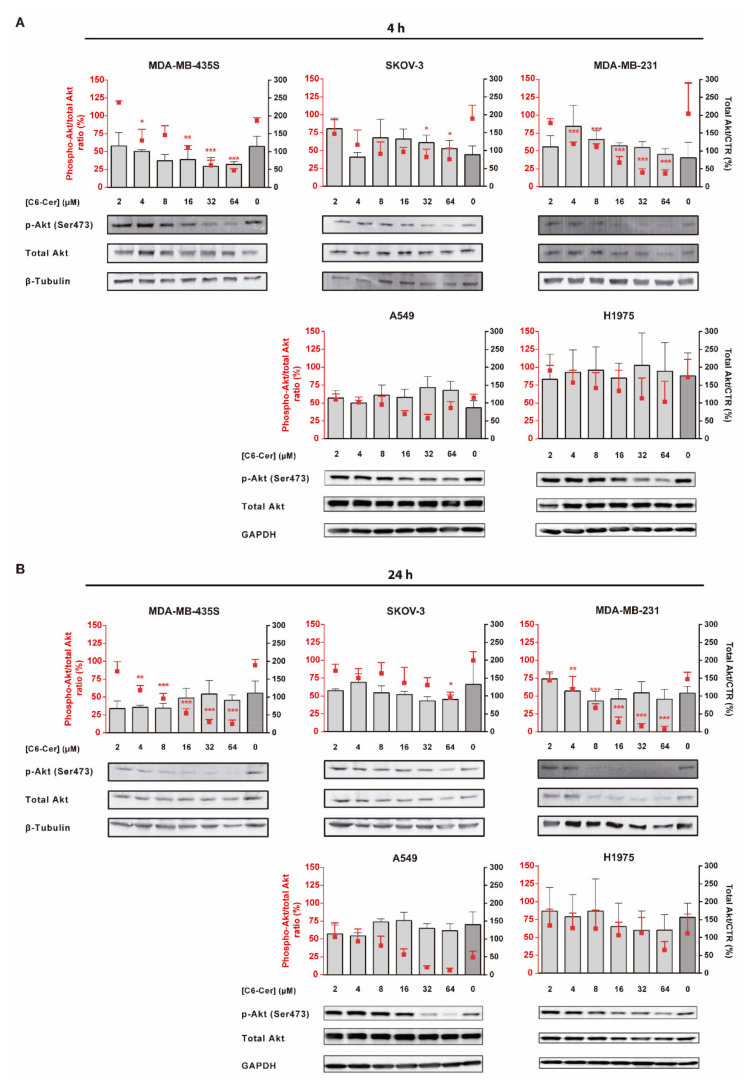
Figure 6.
Evaluation of the modulation of p-Akt protein level by F3 peptide-targeted C6-ceramide liposomes in cancer cell lines of diverse histological origin. MDA-MB-435S, SKOV-3, MDA-MB-231, A549 and H1975 cells were incubated with the indicated concentration of C6-ceramide encapsulated in F3 peptide-targeted liposomes ([F3]L-C6) for (A) 4 h or (B) 24 h at 37 °C. Extracted cellular proteins were analyzed by immunoblotting and band signals for p-Akt (Ser473) and Akt were quantified through densitometry imaging, and the p-Akt/total Akt ratio (red squares) and total Akt/Control (bars) for each condition were calculated. Equal amounts of protein were loaded in each lane. Data represent the mean ± SEM (n = 3; one-way ANOVA with Dunnett’s post-test; * p < 0.05, ** p < 0.01 and *** p < 0.001). Whole Western Blot images can be found in Figure S5.

