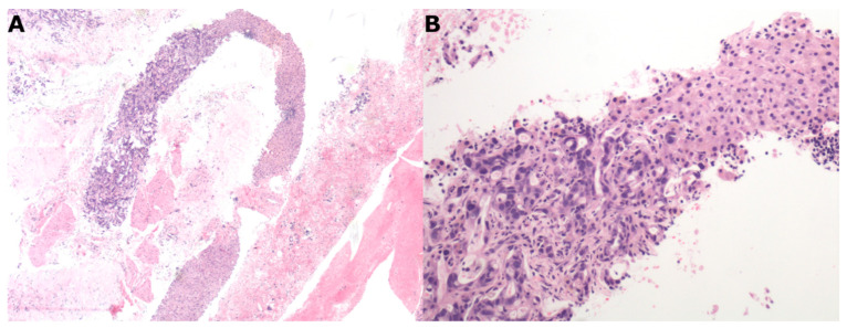
Figure 3.
Liver metastasis from ductal adenocarcinoma of the pancreas. (A) 4 mm needle biopsy of a liver focal lesion with a representative area of adenocarcinoma occupying about 40% of the biopsy (upper left side) (4×, EE). (B) At higher magnification, adenocarcinoma cell aggregates (left side) replacing normal hepatocytes (right side) (20×, EE).

