Figure 4.
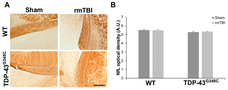
Neurofilament light chain (NfL) expression in the optic tract (OT) of wild-type (WT) and TDP-43G348C mice at 6 months after repetitive mild traumatic brain injury (rmTBI). (A) Representative microphotographs of the OT stained with anti-neurofilament light chain protein. Dashed lines indicate the OT. Scale bar: 200 μm (B) The histogram shows NfL optical density (AU) in the axons of the OT in WT and TDP-43G348C mice with rmTBI and related control groups (Sham). Results are expressed as means ± SEM (N = 3–5).

