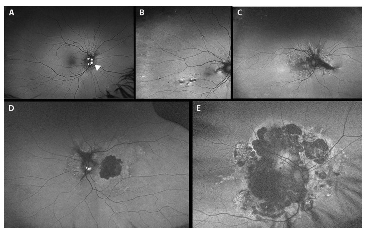
Figure 6.
Fundus auto-fluorescence photographs (A–E) showing: (A) optic disk drusen (white arrowheads); (B) peripheral comet lesions; (C) pattern dystrophy like-changes as hyper/hypo-autofluorescent alterations; (D) pattern dystrophy-like changes with macular atrophy (central hypo-autofluorescence alteration); (E) posterior pole atrophy.

