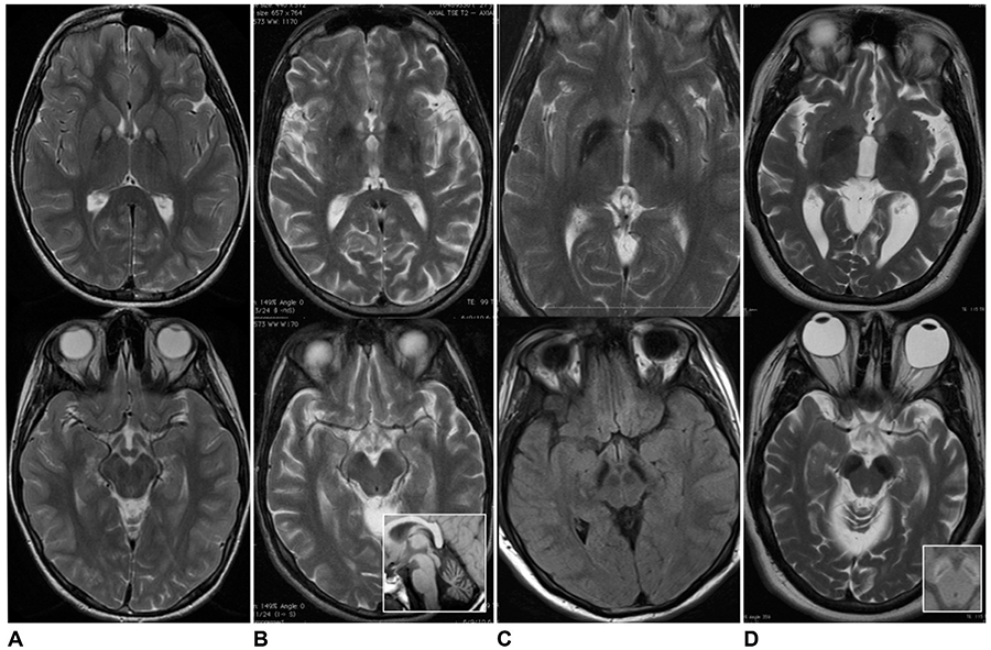
Fig. 19.1.

Magnetic resonance imaging of globus pallidus and substantia nigra in four main forms of neurodegeneration with brain iron accumulation. Axial T2-weighted imaging of globus pallidus (top series) and substantia nigra (bottom series) in (A) pantothenate kinase-associated neurodegeneration; (B) phospholipase A2-associated neurodegeneration (inset shows cerebellar atrophy); (C) mitochondrial membrane protein-associated neurodegeneration; and (D) beta-propeller protein-associated neurodegeneration (inset shows T1 hyperintense “halo” in cerebellar peduncles).
