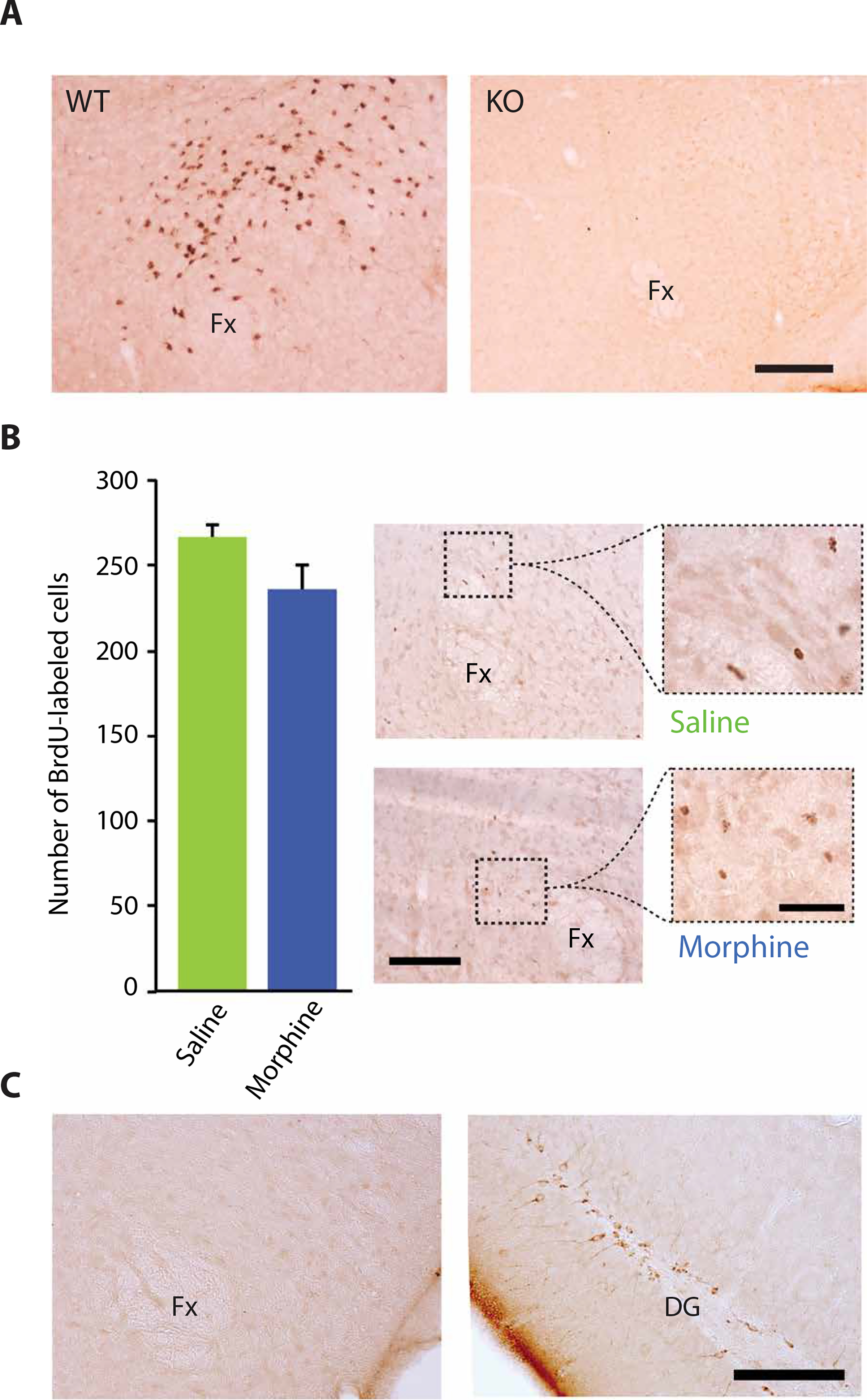
Fig. 4. The increase in hypocretin cell number after morphine administration was not due to neurogenesis.

(A) Morphine administration at 100 mg/kg for 14 days resulted in labeling of hypocretin cells in wild-type (WT) mice, but not in hypocretin knockout (KO) mice, indicating that labeling required the presence of hypocretin in neurons. Scale bar, 200 μm. (B) The increased number of hypocretin neurons after morphine administration was not due to neurogenesis. BrdU labeling to identify new neurons showed no significant increase in the number of new hypocretin neurons after 14 days of morphine (100 mg/kg) treatment of wild-type mice compared to control mice given saline injections. Right: BrdU-labeled cells in the perifornical area of saline-treated (top) and morphine-treated (bottom) animals. Scale bars, 100 and 40 μm. (C) Shown is immunohistochemical staining in the hypothalamus for doublecortin after 14 days of morphine (100 mg/kg) treatment of wild-type mice. Left: Representative photomicrograph shows the absence of doublecortin staining in the hypothalamus of a morphine-treated animal. Right: Representative photomicrograph shows normal doublecortin staining of dentate gyrus (DG) of the same animal. Scale bar, 100 μm.
