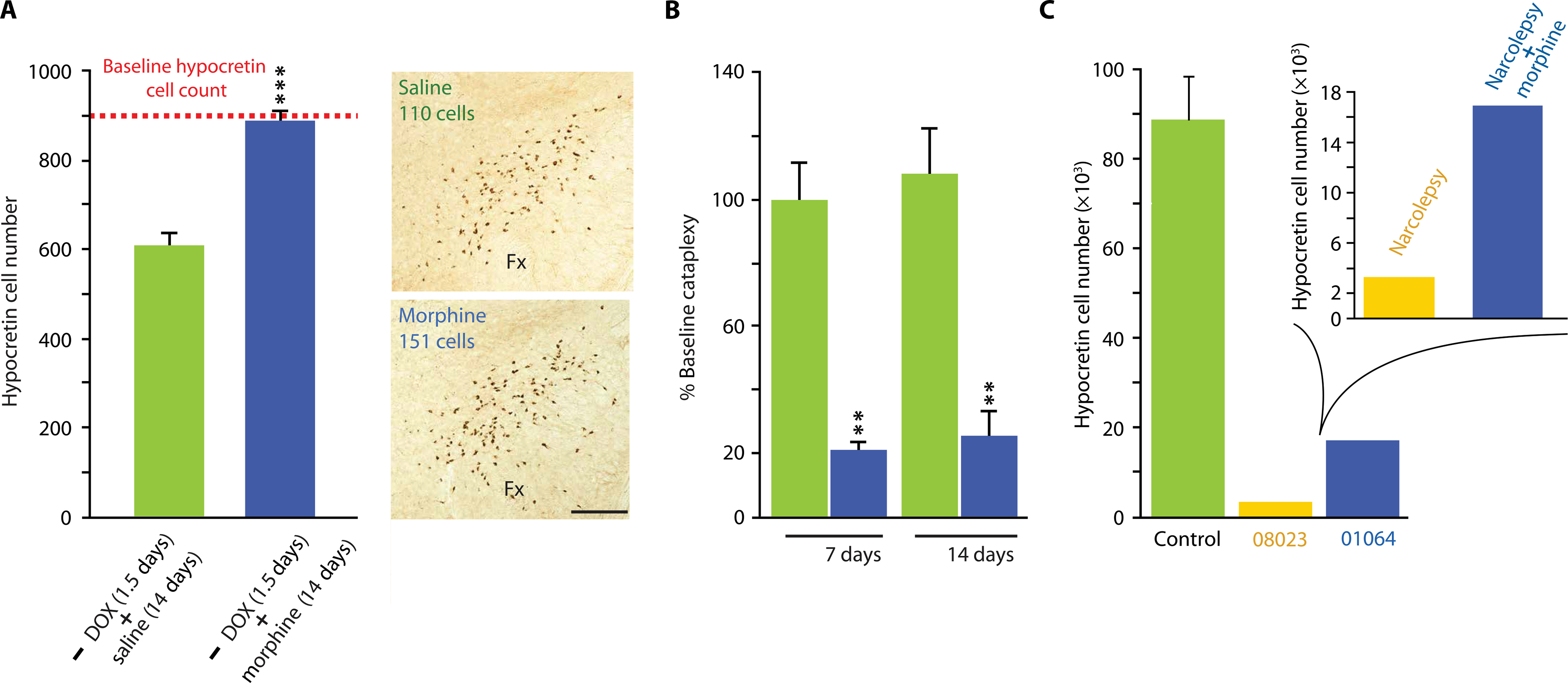
Fig. 6. Reversal of hypocretin cell loss and cataplexy in narcoleptic mice after morphine administration.

(A) The dashed horizontal red line shows the number of hypocretin cells in the brains of narcoleptic DTA mice maintained throughout the experiment on DOX, with 14 days of daily saline injections, followed by sacrifice. The green bar shows the number of hypocretin cells in the brains of DTA mice after DOX withdrawal for 1.5 days, followed by restoration of DOX administration and saline injections for 14 days. A 30% reduction of hypocretin cells relative to control was observed. When daily morphine (100 mg/kg) injections were given instead of saline for 14 days, the number of detected hypocretin cells was restored to baseline (blue bar). This difference was significant [***P = 0.003, t = 6.31, df = 4 (t test)]. Photomicrographs on the right show representative examples of hypocretin labeling in brains of DTA mice treated with saline (top) or morphine (bottom). Scale bar, 200 μm. (B) Morphine administration (blue) to DTA mice decreased cataplexy after 1 and 2 weeks of administration relative to control DTA mice receiving saline injections (green). Treatment effect: F1,6 = 148.4, P = 0.0001 (ANOVA). Changes after saline administration were not significant. Post hoc comparisons with Bonferroni correction revealed a significant difference at P < 0.01 between the saline-treated control DTA mice and the morphine-treated DTA mice at both 1 and 2 weeks of treatment. (C) Immunohistochemical staining shows that brain tissue from a human narcoleptic patient with cataplexy (01064, blue) treated for a long period with morphine has a higher number of hypocretin cells than does a case control narcoleptic patient with cataplexy (08023, yellow) not treated with morphine. The hypocretin cell counts in brain tissue from three control patients without narcolepsy or other identified neurological disorders are shown in green. The patients’ brains were willed to the Netherlands Brain Bank and preserved and analyzed using the same techniques (Table 1).
