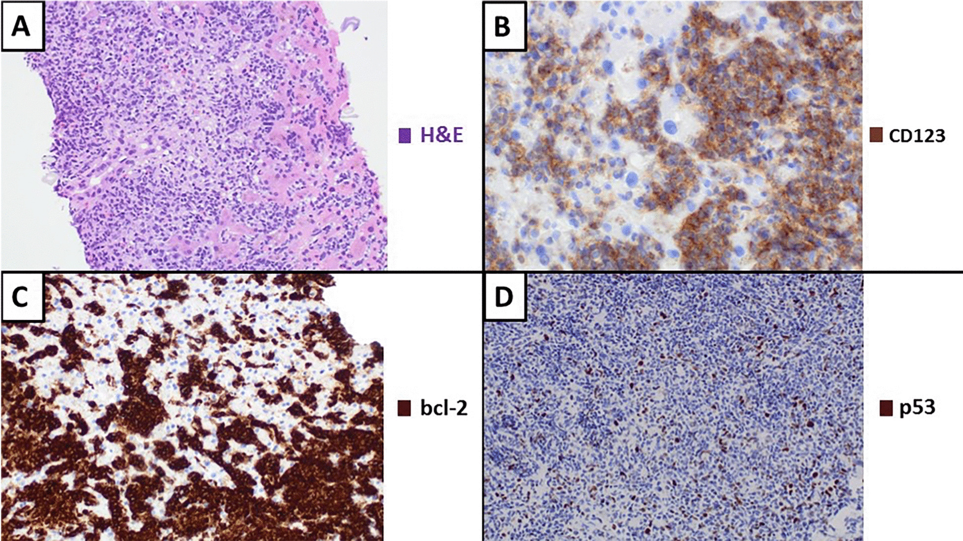
Fig. 3.

Histological sections of the liver biopsy showed a nodular infiltrate of blast cells, with ample cytoplasm, irregular nuclei and small nucleoli. There were occasional small lymphocytes intermingled with neoplastic cells. Tumour cells were diffusely positive for TdT, CD43 and CD56, as well as CD123. Only heterogeneous expression of CD4 was observed. A Nodular infiltration of the liver by blast cells. B Neoplastic cells show diffuse immunoreactivity for CD123. C Positive immunoreactivity for bcl-2. D Occasional intense positivity for p53 (less than 3%)
