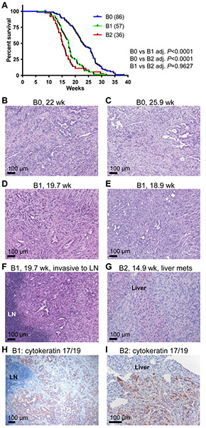
Figure 5. HMMRΔexon8-16 shortened the survival of p48-Cre; LSL-KRASG12D; p53lox/+ mice.
(A) Kaplan-Meier survival curve for B0: 86 mice, B1: 57 mice, and B2: 36 mice. Tukey adjusted P values from pairwise log-rank test were shown. (B-E) H&E stain of invasive PDAC from (B) a 22-week-old B0: p48-Cre; LSL-KRASG12D; p53lox/+ mouse, (C) a 25.9-week-old B0: p48-Cre; LSL-KRASG12D; p53lox/+ mouse, (D) a 19.7-week-old B1: p48-Cre; LSL-KRASG12D; p53lox/+; HMMRΔexon8-16/WT mouse, and (E) a 18.9-week-old B1: p48-Cre; LSL-KRASG12D; p53lox/+; HMMRΔexon8-16/WT mouse. (F) H&E stain of a peripancreatic lymph node (LN) invaded by PDAC from the same animal in (D). (G) H&E stain of liver metastasis of PDAC from a 14.9-week-old B2: p48-Cre; LSL-KRASG12D; p53lox/+; HMMRΔexon8-16/Δexon8-16 mouse. (H, I) Immunohistochemical staining of cytokeratin 17/19 of the lymph node (LN) invasion (H) and liver metastasis (I).

