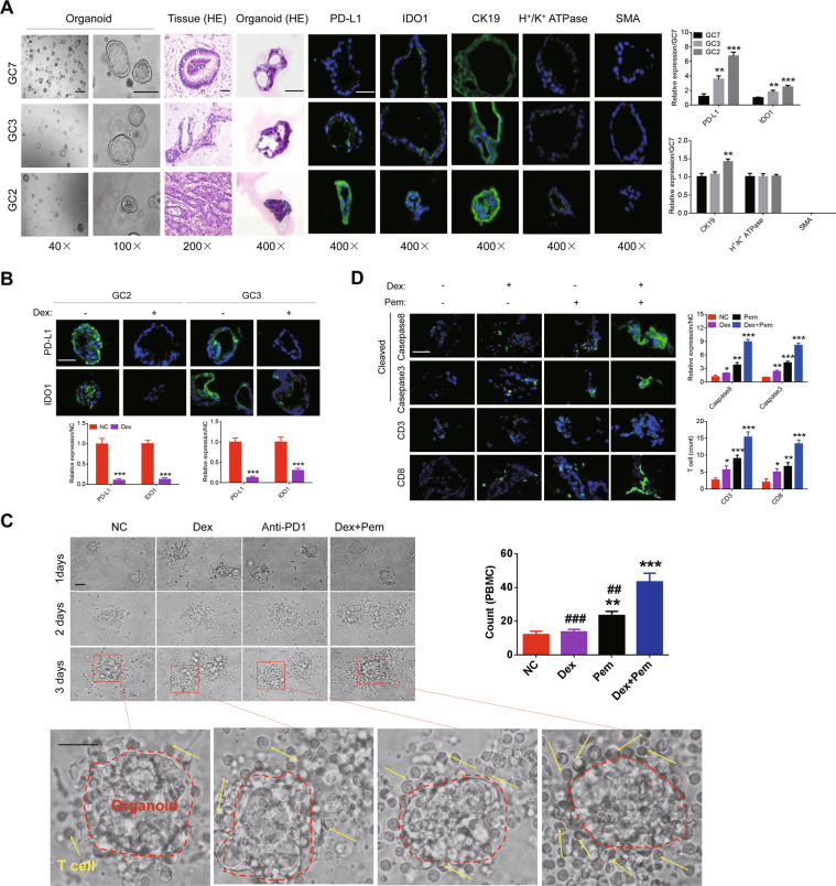
Fig. 5. Dexamethasone increases efficacy of ICIs in the ex vivo organoid models.
A Organoids were constructed from fresh GC tissues (left; 40× and 100×); and HE staining of organoids and organoids-derived tissues was performed (200× and 400×). The expression of PD-L1, IDO1, CK19, H+/K+ ATPase, and SMA on organoids was detected by immunofluorescence. B Expression of PD-L1 and IDO1 in organoids (GC3 and GC2) treated with low-dose of dexamethasone (200×). C After pretreatment with 50 nM dexamethasone for 72 h, organoids were co-cultivated with PBMCs at an effector/ target ratio of 5:1, and then incubated with dexamethasone (10 nM) or PD1 inhibitor (10 μg/ml) for 48 h. Bright-field microscopy image showed the PBMCs surrounding organoids (200× and 400×); D The cells surrounding around organoid were evaluated for the expression of CD3, CD8, caspase3, and caspase8 by immunofluorescence (400×). “*” indicates comparison with NC; “#” indicates comparison with “dexamethasone and pembrolizumab”. The results were repeated three times and shown as the mean ± SD.

