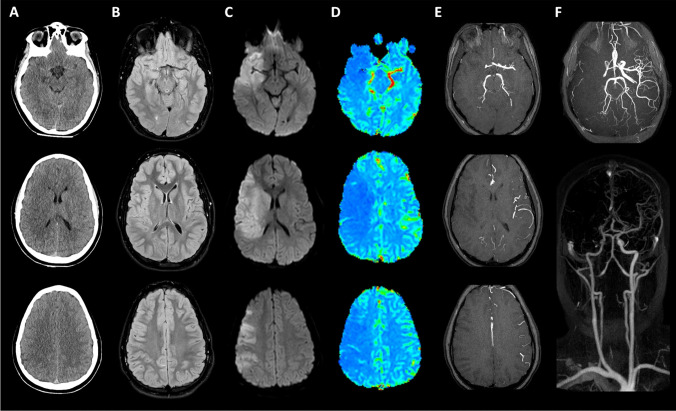
Fig. 1.
Early CT scan and fast MR study at admission. First CT scan (A) showed only slight hypoattenuation and obscuration of the head of the caudate nucleus as well as a loss of gray/white matter definition in the lateral margins of the right insula, findings suggestive of “early signs” of ischemic stroke; slight hyperattenuating band on the right middle cerebral artery (MCA) was also observed, due to intravascular occluding thrombus. Similarly, MR FLAIR images (B) were nearly normal, with only subtle hyperintensity along with the cortical insular ribbon. Instead, diffusion-weighted [b 1000 s/mm2] images (C) clearly showed acute stroke–induced cytotoxic edema in the right MCA territory; the discrepancy between DWI and FLAIR images was suggestive of an early stage of stroke. The MR perfusion CBF map (D) demonstrated a large wedge-shaped area of significantly reduced perfusion in the same region, also corresponding to a lack of vascular representation on MRA TOF3D MIP reconstructions (E). MR-angiography (F) performed with TOF3D, and contrast-enhanced techniques confirmed the occlusion of M1 segment of the right MCA

