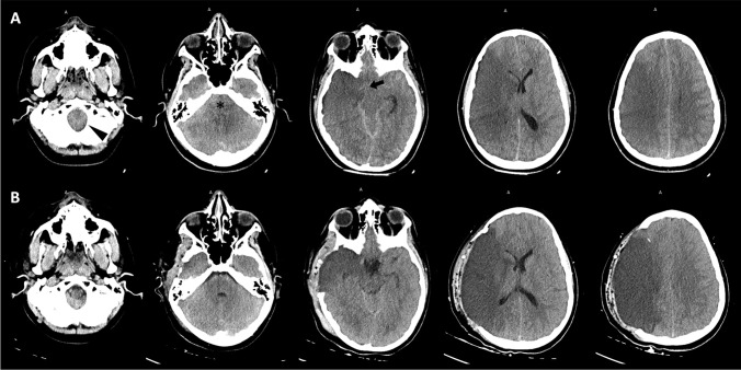
Fig. 2.
Brain CT monitoring. Post-endovascular treatment CT scan (A), performed after the onset of anisocoria, demonstrated extensive swelling in the right hemisphere, right transtentorial uncal herniation (black arrow), effacement of the CSF spaces in the posterior fossa (asterisk), incipient tonsillar herniation (arrowhead) and leftward midline shift due to malignant cerebral edema. These compressive findings were improved on CT images (B) obtained after DHC

