Fig. 1.
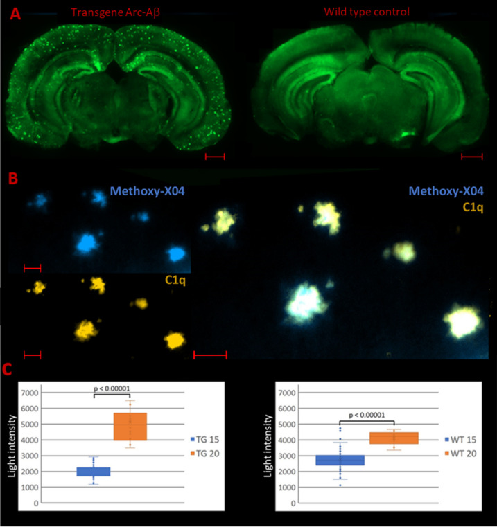
C1q staining in transgenic C57BL/6 Arc-Aβ mice and corresponding wild type control animals. a Immunofluorescence images of PFA fixed free-floating stained brain sections using the monoclonal anti-C1q antibody Abcam 182541: C1q staining (green) is distributed over the entire brain. C1q plaques only occur in the transgenic Alzheimer’s disease mouse model Arc-Aβ (scale bars: 1000 µm). b Colocalization of C1q and β-amyloid plaques: C1q depositions (orange) continuously overlap with Methoxy-X04 stained β-amyloid plaque accumulations (scale bars: 50 µm). c Light intensity measurements of C1q stained brain sections: C1q expression within the dentate gyrus differs significantly between 15- and 20-months-old animals (Mann–Whitney U test: p < 0.00001). This effect is even more pronounced in the transgenic mouse model (TG transgenic, WT wild type)

