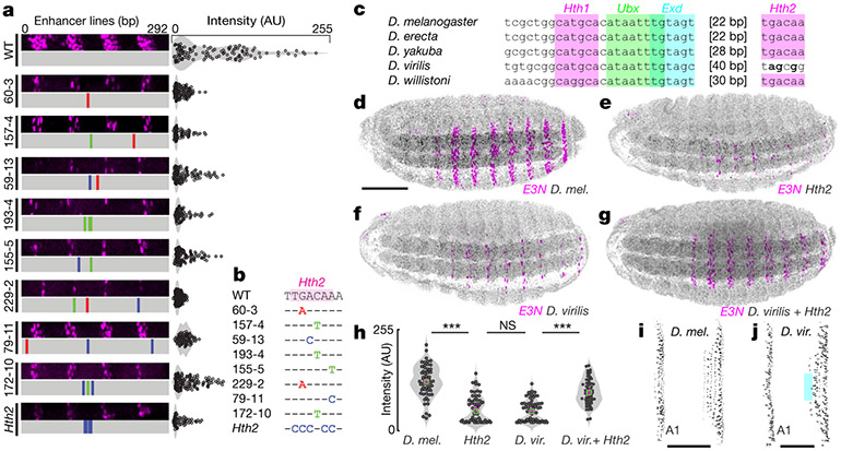
Fig. 2 ∣. Mutational scanning identifies a Hth binding site associated with a changed evolutionary phenotype.
a, Ventral abdominal segments from lines with mutations in an Hth2 binding site (left) and individual cell-staining intensities (right). b, Sequences of Hth2 binding site in lines tested. c, Conservation of E3N in Drosophila, highlighting conserved binding sites (coloured areas). d–g, Embryos bearing E3N::lacZ constructs in a D. melanogaster background, stained with anti-β-galactosidase. d, D. melanogaster (D. mel.) wild-type E3N::lacZ reporter construct. e, D. melanogaster E3N::lacZ reporter construct with a mutated Hth2 site. f, Drosophila virilis E3N::lacZ reporter construct. g, D. virilis E3N::lacZ reporter construct with rescued Hth2 site (5′-TGACAA). h, Single-cell quantification of staining intensities in embryos bearing indicated constructs. D. vir., D. virilis. Magenta crosses, mean; green squares, median. Two-tailed t-test; ***P < 0.01, NS not significant (n = 50, 10 embryos). Left to right: P < 0.01, P = 0.34, P < 0.01. i, j, Cuticle preparations for D. melanogaster (k) and D. virilis (l) showing ventral regions. Scale bars, 50 μm. Teal rectangle highlights trichomes that are absent in D. virilis. f, Scale bar, 100 μm; embryos in d–g matched for scale.

