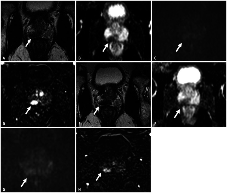
Fig. 4. A 62-year-old male presented with an elevated serum PSA of 5.9 ng/mL.
mpMRI was performed as a first-line investigation.
A. T2-weighted imaging shows a focal lesion in the peripheral zone at the right base (arrow). B. ADC map shows marked hypointensity (arrow). C. DWI shows mild hyperintensity (arrow). D. DCE imaging shows intense early enhancement (arrow). The lesion was graded as Prostate Imaging-Reporting and Data System 4. Systematic transrectal ultrasound biopsy showed a Gleason 3 + 3 cancer. Initial MRI-ultrasound fusion targeted biopsy was negative for malignancy. A repeat targeted biopsy was also negative, and the patient was enrolled for active surveillance. Repeat mpMRI was performed after 1 year before confirmatory biopsy. Repeat PSA was 5.7 ng/mL (stable). E. T2-weighted imaging shows a relatively stable size of the hypointense lesion in the right midgland peripheral zone (arrow). F, G. ADC map and DWI show similar findings as before (arrows). H. DCE imaging shows intense early enhancement (arrow). The lesion was deemed stable (Prostate Cancer Radiological Estimation of Change in Sequential Evaluation score 3), and targeted biopsy was not recommended. Patients underwent systematic confirmatory biopsy that showed a Gleason 3 + 3 cancer. Subsequent mpMRI before surveillance systematic biopsy at 2 years from initial diagnosis continued to show stable imaging findings. ADC = apparent diffusion coefficient, DCE = dynamic contrast-enhanced, DWI = diffusion-weighted imaging, mpMRI = multiparametric MRI, PSA = prostate-specific antigen

