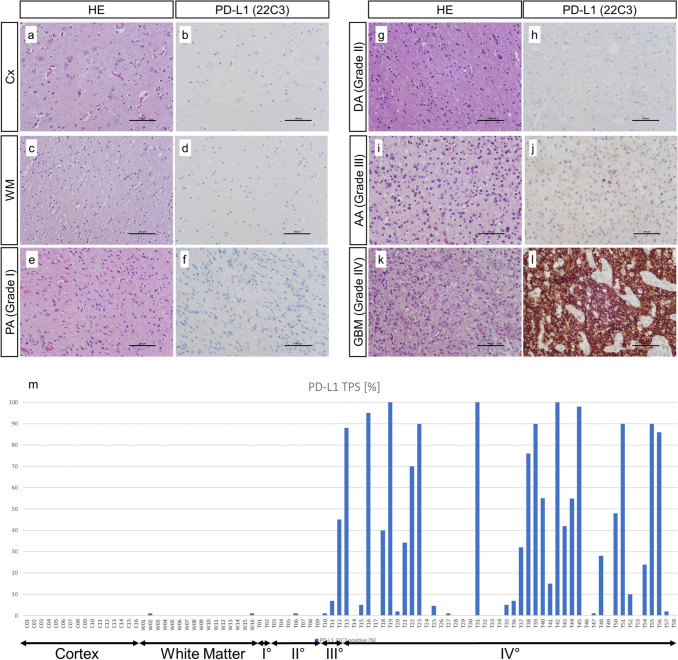
Fig. 1.
PD-L1 expression in healthy brain tissue and glioma. Analysis of 90 tissue samples showed no PD-L1 expression in healthy cortex (a, b) and white matter regions (c, d). There was no noteworthy PD-L1 expression in low grade glioma, i.e., WHO grade I pilocytic astrocytoma (e, f) and diffuse astrocytoma (g, h). In high grade glioma there was an uneven PD-L1 expression with strong intertumoral heterogeneity in WHO grade III anaplastic astrocytoma (i, j) and glioblastoma (k, l). Distinct PD-L1 TPS scores of all 90 analyzed samples is presented in m. CX cortex, WM white matter, PA pilocytic astrocytoma, DA diffuse astrocytoma, AA anaplastic astrocytoma, GBM glioblastoma. a–l Scale bar: 100 µm

