
FIG. 2.
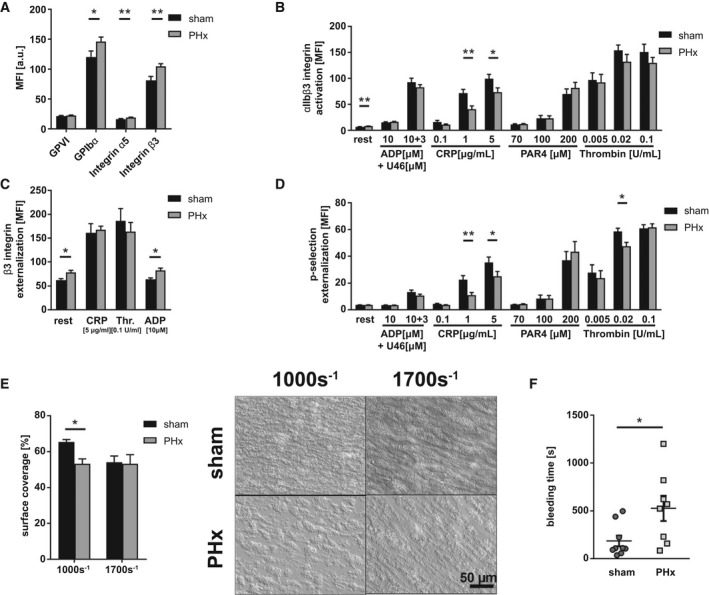
Platelet activation defects and reduced thrombus formation lead to bleeding complications 3 days after PHx. (A) Expression of indicated glycoproteins on the surface of platelets measured by MFI in flow cytometric analysis (n = 13 sham, 18 PHx). (B) Externalization of P‐selectin on the platelet surface with indicated agonists (n = 9 sham, 12 PHx). (C) Externalization of the β3 integrin subunit upon platelet stimulation (n = 8 sham, 9 PHx). (D) Activation of ⍺IIbβ3 integrin on the platelet surface with indicated agonists (n = 9 sham, 12 PHx). (E) Thrombus formation on a collagen matrix under arterial shear rates with representative pictures (1.000 and 1.700 s−1; n = 3 sham, 4 PHx). (F) In vivo analysis of hemostatic dysfunction by tail bleeding time (n = 9 sham, 8 PHx). Depicted are mean values + SEM; *P < 0.05; **P < 0.01, using paired Student t test. Abbreviations: a.u., arbitray units; MFI, mean fluorescence intensity; Thr., thrombin.