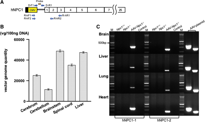
Figure 2.
Vector genome distribution and expression of hNPC1. (A) Location of the primers and probe. The open vertical rectangles represent coding exons. The vector genome DNA was amplified using the primer set of D-F1 and D-R1. The hNPC1 mRNA was amplified using the two primer sets of R-hF and R-hR. (B) Quantitation of the vector genome by qPCR. The vector genome was detected in the broad area of CNS, including the brain stem and spinal cord. It was also detected in the liver. (C) Detection of transgene-specific mRNA in the brain, liver, lung, and heart. The expression of transgene-specific hNPC1 was detected only in the AAV-treated Npc1−/− mice, whereas no fragments were amplified in the untreated Npc1+/+ or Npc1−/− mice. We used the AAV9/3-CMV-hNPC1 plasmid as a positive control. vg/100 ng DNA, vector genomes per 100 ng DNA; M, molecular marker; Control, control without template cDNA. CNS, central nervous system; qPCR, quantitative PCR.

