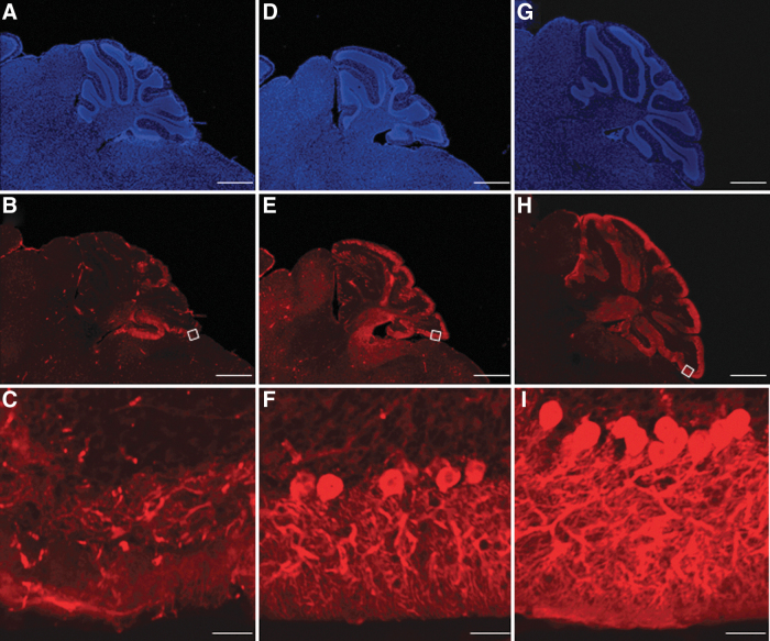
Figure 4.
Preservation of Purkinje cells. The cerebella of untreated Npc1−/− (A–C), AAV-treated Npc1−/− (D–F), and untreated Npc1+/+ mice (G–I) at week 11 stained with anti-calbindin antibody and Hoechst. While there were only a few Purkinje cells left in the untreated-Npc1−/− mice, far more cells were observed in the AAV-treated Npc1−/− mice. Scale bar = 500 μm (A, B, D, E, G, H), 50 μm (C, F, I).

