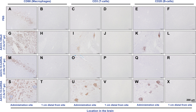
Figure 5.
IHC assessment of inflammatory cells at the sites of administration of AAVrh.10hARSA, AAVrh.10Null, and PBS. Staining of the brain coronal sections from 1, 13, and 26 weeks NHP was done to assess the MRI ROIs and microscopic findings of concern to identify the types of inflammatory cells. The slides were stained by IHC for CD68 (microglial cells and monocyte-derived macrophages), CD3 (T cells), and CD20 (B cells). The resulting brain immunohistochemical staining is shown for the vector administration site and 1 cm distal from the administered site for PBS (A–F), AAVrh.10Null (G–L), low dose (M–R), and high dose (S–X) AAVrh.10hARSA groups. Black scale bar = 200 μm. IHC, immunohistochemistry.

