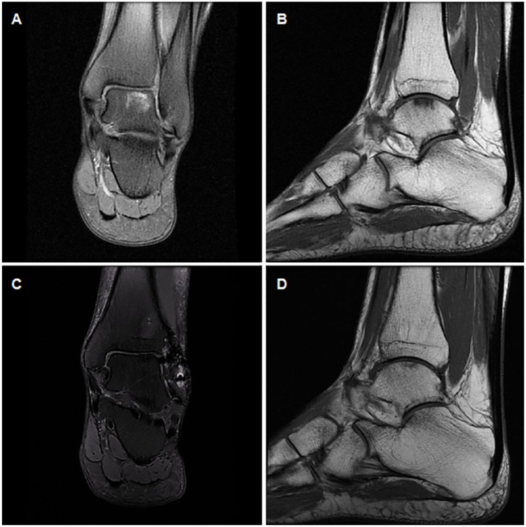
Figure 5.
Coronal and sagittal magnetic resonance imaging (MRI) from a representative case of an International Cartilage Repair Society (ICRS) grade 3 osteochondral lesion located on the talus medial area (144 cm2, American Orthopaedic Foot & Ankle Society [AOFAS] score 48) (A, B). At 24-month follow-up, defect was filled by a smooth tissue very similar to hyaline cartilage (AOFAS score 90; magnetic resonance observation of cartilage repair tissue [MOCART] score 84 (C, D). The persistence of abnormalities in subchondral bone is noteworthy.

