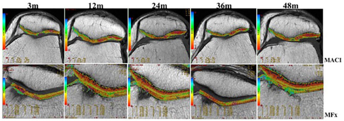
Figure 6.
Quantitative assessment of repaired cartilage by T2-mapping. Invasive imaging method that provides quantitative values that represents the quality of the repair tissue. Similar behavior was observed at 3 postoperative months with the presence of a high signal intensity in both groups. By 12 and 24 months, treated defects in both techniques had well-defined surface and intermediate signal intensity with more closed values to native cartilage without significant difference between experimental groups. T2-mapping values improved significantly over time in MACI compared to MF at 48 months (P < 0.05). At most MACI sites (above), the chondral defect was completely filled with homogeneous tissue to the expected level of the adjacent articular cartilage.

