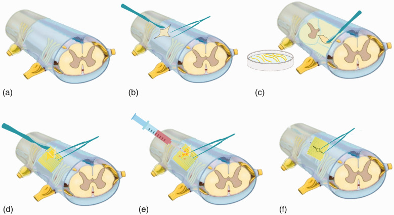
Figure 1.
Images showing the spinal cord surgery performed in the monkeys. (a) Unmanipulated spinal cord after total laminectomy at the T8 level. (b) The dura mater was opened longitudinally at the midline to uncover the spinal cord. (c) The sural nerve was harvested, cut into several segments, and placed in Hank’s balanced salt solution. (d) Sural nerve segments were extracted and used to fill the space of the spinal cord stumps. (e) Acidic fibroblast growth factor was infused into the grafted site. (f) The dura mater was sutured.

