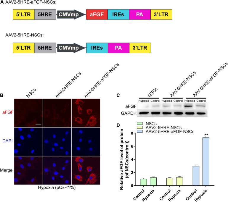
FIGURE 1.
The preparation and identification of AAV–5HRE–aFGF–NSCs. (A) The schematic diagrams of the vector construction: AAV–5HRE–aFGF and AAV–5HRE. (B) The immunofluorescence shows the expression of aFGF (red) and DAPI (blue) in different groups. Magnification: 40×; Scale: 50 μm. (C) Western blotting showing the expression of aFGF in each group. (D) The quantitative analysis of aFGF protein expression. **P < 0.01. Data are represented as mean ± SD (n = 6). AAV, adeno-associated virus; 5HRE, five hypoxia-responsive elements; aFGF, acidic fibroblast growth factor; NSCs, neural stem cells.

