Figure 3.
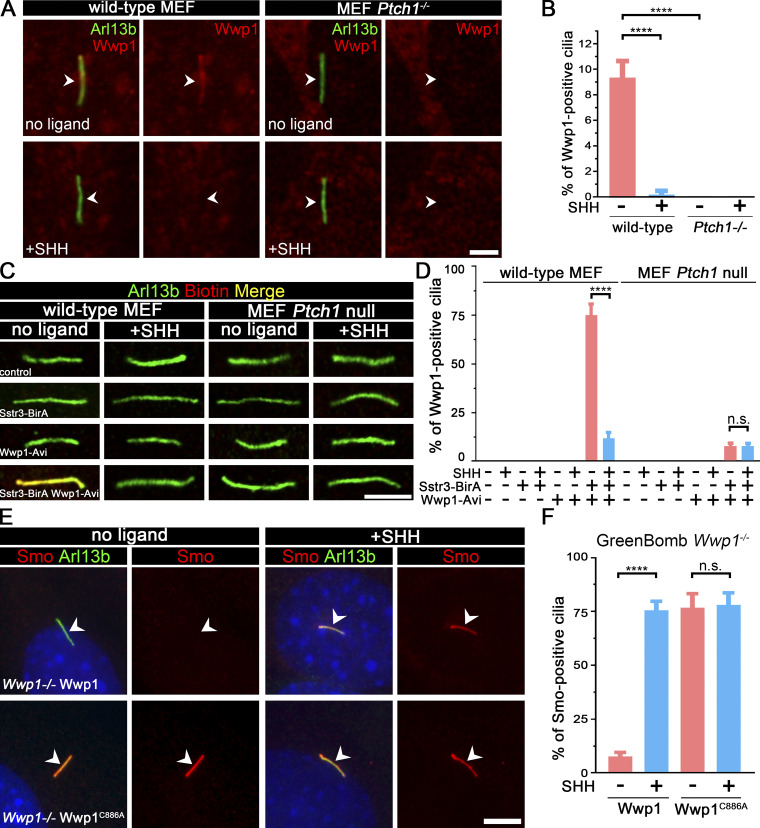
Wwp1 localizes in cilia, and its E3 ligase activity is required for the ciliary removal of Smo.(A) WT MEF or MEF Ptch1-null cells expressing Wwp1-Flag were stained for Wwp1 (Flag, red) and cilia (Arl13b, green, arrowheads) with or without exposure to SHH-conditioned medium. Scale bar, 2 microns. (B) Quantification of the percentage of Wwp1-positive cilia from A. n = 6 replicates with at least 200 cilia counted each time. ****, P < 0.0001 by two-way ANOVA. Error bars indicate SD. (C) WT MEF or MEF Ptch1-null cells expressing Sstr3-BirA and Wwp1-Avi were stained for Wwp1 (biotin, red) and cilia (Arl13b, green) with and without SHH. Scale bar, 3 microns. (D) Quantification of the percentage of Wwp1-positive cilia from C. n = 6 repeats with 200 cilia counted per experiment. ****, P < 0.0001 by two-way ANOVA. Error bars indicate SD. (E) Wwp1 mutant cells transfected with Wwp1-Flag show rescue of ciliary Smo levels. Wwp1C886A is enzymatically dead, indicating that E3 activity is required for rescue. Arrowheads mark cilia. (F) Quantitation of Smo-positive cilia described in E. n = 6 repeats with 200 cilia counted per experiment. ****, P < 0.0001 as compared with serum-starved cells by two-way ANOVA. Error bars indicate SD.

