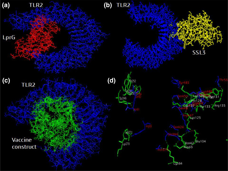
Fig. 7.
Molecular docking of the designed and validated vaccine construct with TLR2 immune receptor. a TLR2 agonist LprG protein (red color) and c modeled vaccine construct (green color) binds to the active site groove of TLR2 receptor (blue color). b The negative control SSL3 protein (yellow color) binds randomly to TLR2 receptor (blue color). d The hydrogen bonds formed between the active site residues of TLR2 molecule labeled in red with the vaccine construct residues labeled in white

