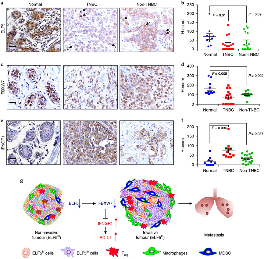
Fig. 8 ∣. Compared with normal breast cells, tumour cells have lower expression of the proteins ELF5 and FBXW7 and higher expression of IFNGR1.
a-f, IHC of TNBC and non-TNBC tumours along with adjacent normal tissue was performed using antibodies against ELF5 (a and b), FBXW7 (c and d) and IFNGR1 (e and f). Images (a, c and e) and graphical representations (b, d and f) show high ELF5 (n = 10 for normal; n = 16 for TNBC; n = 17 for non-TNBC) and high FBXW7 staining (n = 12 for normal; n = 14 for TNBC; n = 11 for non-TNBC) in normal tissues compared with TNBC and non-TNBC tissues, and higher expression of IFNGR1 in TNBC and non-TNBC tumours compared with adjacent normal tissues (n = 10 for normal; n = 12 for TNBC; n = 18 for non-TNBC tissue). In all instances, n denotes independent biological samples. Nuclear localization of ELF5 and FBXW7 was considered positive in tumour tissues. Quantification of H-scores (intensity × abundance) showed a significant difference in ELF5 and FBXW7 expression among normal and TNBC tissues and significantly higher expression of IFNGR1 in TNBC and non-TNBC compared with adjacent normal tissues. The arrows indicate nuclear ELF5 in tumour tissues. Cytoplasmic/membrane staining of IFNGR1 was scored as positive in tissues by IHC. Statistical significance was determined by Mann–Whitney U-test in b, d and f. The data are presented as means ± s.e.m. Scale bars: 40 μm (a, c and e). g, Schematic showing that low ELF5 and FBXW7 in TNBC tumour cells leads to stabilization of IFNGR1, resulting in increased PD-L1 expression. This increases invasive properties in TNBC tumour cells, followed by increased metastasis. MDSC, myeloid-derived suppressor cell.

