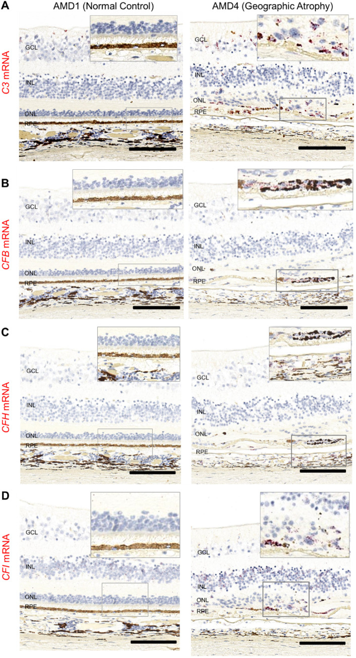
Figure 3.
Spatial distribution of complement C3, CFB, CFH, and CFI mRNAs in macular area AMD1 and AMD4 macula. Donor eyes with AMD1–4 were evaluated for complement gene expression using RNAscope technology on paraffin sections. In representative examples of an AMD1 and an AMD4 macula, we identify changes in mRNA distribution (red): (A) C3 mRNA expression present in the GCL and INL of AMD1 macula but not in outer retina (left image). In addition to inner retina C3 expression, there is a strong C3 positive signal in the atrophic area of the AMD4 donor (right image). (B) CFB mRNA positive signal is present in the GCL and INL of AMD1 macula, but not in outer retina (left). In addition to staining of the GCL and INL, in the GA lesion area there is a strong CFB positive signal (right). (C) CFH mRNA expression is present in RPE, retinal and choroidal vessels of both AMD1 and AMD4 macula. In the atrophic lesion, CFH positive signal is prominent in a patch of abnormal-appearing RPE. (D) CFI mRNA expression is present in GCL, INL, and also in RPE and choroid of AMD1 and AMD4 macula. In the atrophic area of AMD4, there is readily detectable CFI expression in the subretinal space. Total number of eyes examined: AMD1 N = 3, AMD4 N = 4. Scale bar: 100 µm.

