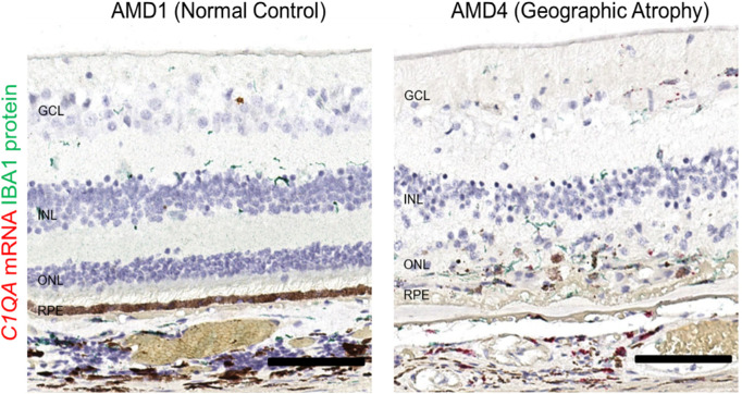
Figure 5.
Spatial distribution of complement classical pathway gene expression C1QA relative to IBA1+ cells in AMD1 and atrophic lesion region of AMD4 macula. (A). In an AMD1 macula, very few C1QA mRNA positive signals (red) were present and were exclusively in IBA1+ cells (green) (left image). Numerous C1QA mRNA expressing amoeboid-shaped IBA1+ cells were observed in atrophic lesion of an AMD4 eye (right image). These cells contained several copies of C1QA mRNA molecules. Total number of eyes examined: AMD1 N = 3, AMD4 N = 4. Scale bar: 100 µm.

