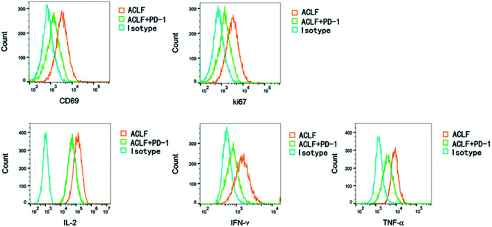
Fig. 7. When PD-1/PD-L1 was activated, the cell viability (CD69) (MFI: 917±43 vs. 1,723±143, p<0.001] and proliferative ability (Ki67) (MFI: 940±71 vs. 1,737±139, p<0.001) of CD8+ T lymphocytes in patients in the ACLF+PD-1 group were lower than those in the ACLF group.
The levels of IL-2 (MFI: 64,267±3,644 vs. 150,587±9,157, p<0.001), IFN-γ (MFI: 1,307±96 vs. 1,737±161, p=0.031), TNF-α (MFI: 2,100±119 vs. 4,050±242, p<0.001) were lower than those in the ACLF group. ACLF, acute-on-chronic liver failure; IFN-γ, interferon-gamma; MFI, mean fluorescence intensity; PD-1, programmed cell death-1; PD-L1/2, programmed cell death 1-ligand 1/2; TNF-α, tumor necrosis factor-alpha.

