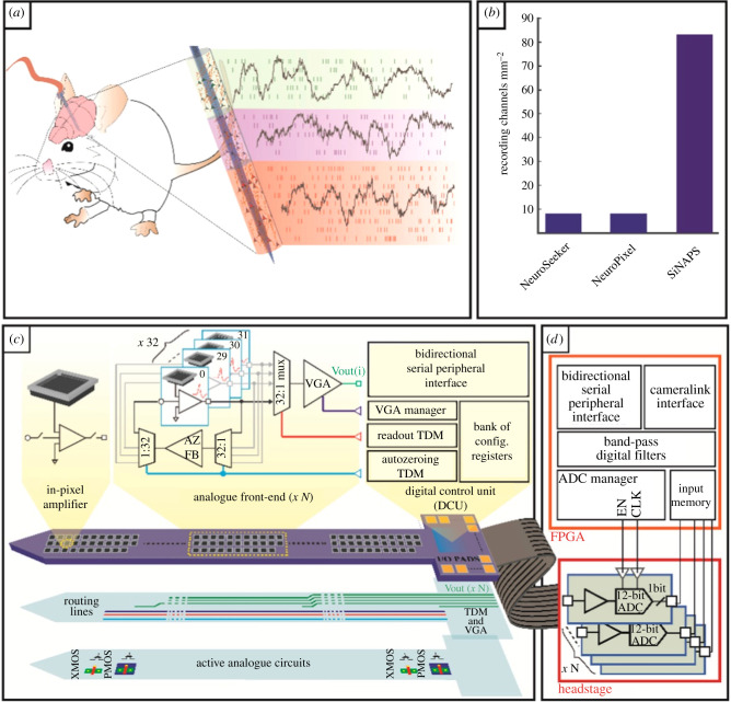
Figure 2.
Architecture of the SiNAPS probe and of the recording system. (a) Implantable CMOS (complementary metal-oxide semiconductor) probes with dense electrode arrays can record broad-band bioelectrical signals across brain circuits with sub-millisecond and single-neuron resolutions. (b) Comparison of the integration potential of simultaneously recording electrodes (i.e. channels) per total silicon area (i.e. shaft and base of the probe) for different architectures proposed in the literature. SiNAPS probes achieve a number of effectively recording channels per unit of silicon area that is one order of magnitude larger than other presently available CMOS architectures and the NeuroPixels. (c,d) Schematics of the circuit architecture for the SiNAPS probe (c) and its acquisition system providing simultaneous neural recordings from the entire electrode array (d). Each electrode-pixel features an electrode and a small area DC-coupled in-pixel circuit for local amplification and low-pass filtering. A probe integrates multiple instances of the same low-area and low-power analogue front-end module of 32 electrode-pixels that are read out in a time-division multiplexed fashion. The on-probe digital control unit (DCU) provides the timing signals required for correct circuit operation and implements a bidirectional serial peripheral interface (SPI) for device configuration. A field-programmable gate array (FPGA)-based acquisition unit generates timing signals for the analog to digital converters (ADCs) and provides a camera link standard connection with a PC for data storage and online visualization [38]. TDM, time division multiplexing; VGA, video graphics array. (Online version in colour.)

