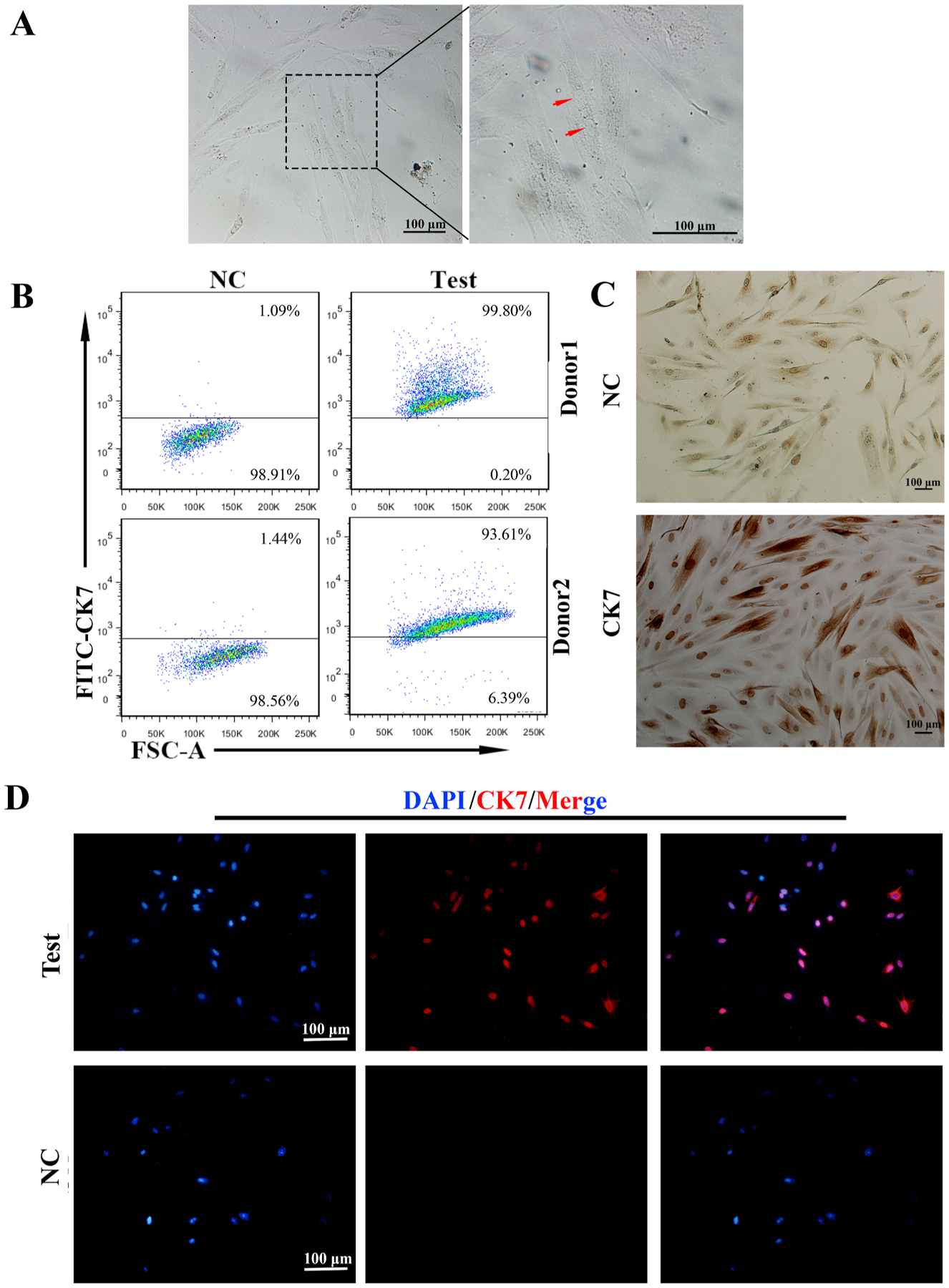
Fig. 1.

The preparation of highly purified primary trophoblasts.
A: The images of primary trophoblasts taken with different magnification. The left and middle ones showed the irregular and polygon cells in the field with 3 days cultures (i). The right one (iii) revealed three nuclei found in one cell (arrow) around 10 days cultures.B: The purity of primary trophoblasts by flow cytometry (FCM). The CK7+cells were trophoblasts, which reached 93% to 99% of cell populations.C: CK7 localization in primary trophoblasts by immunocytochemistry. (i)The upper panel was negative control. (ii)Almost all of the cells were stained with CK7 in these fields (lower panel). CK7 was localized in the cytoplasm and nucleus, especially deeply stained in the nucleus. Scale bars: 100 μm.D: Most of the primary trophoblast cells expressed trophoblast specific marker, CK7 in the field (upper panel). CK7 was mainly localized in the nucleus by Immunofluorescence (IF) staining. The lower panel was negative control (NC). Scale bars: 100 μm.
