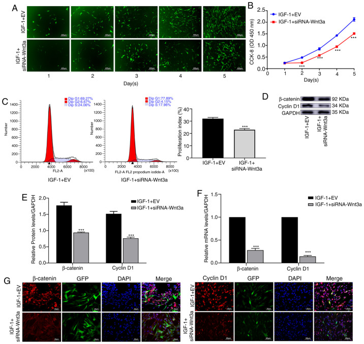
Figure 4.
By inhibiting the Wnt/β-catenin pathway, IGF-1 reduces the proliferation of BMSCs. BMSCs were transfected with empty vector or the siRNA-Wnt3a gene. Then, the cells were treated with IGF-1 (80 ng/ml). (A) The proliferation of the cells for 5 consecutive days using a fluorescence microscope. (B) CCK-8 was used to detect the proliferation of BMSCs. (C) Cell cycle detection assays were used to detect the proliferation of BMSCs. (D and E) β-Catenin and cyclin D1 protein expression was determined by western blotting. (F) The relative mRNA expression of β-catenin and cyclin D1 was evaluated by quantitative polymerase chain reaction. (G) Immunofluorescence was used to detect the expression of β-catenin and cyclin D1 in BMSCs. ***P<0.001 vs. IGF-1 + EV. IGF, insulin growth factor; BMSCs, bone marrow mesenchymal stem cells; CCK-8, Cell Counting Kit-8; si, small interfering; EV, empty vector.

