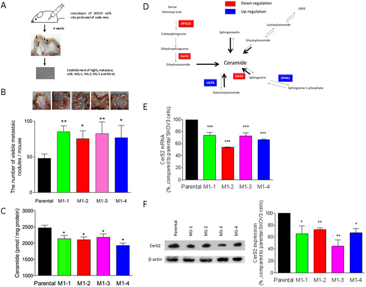
Figure 1. In vivo functional screening and characterization of metastasis-prone sublines.
(A) For in vivo functional screening, five million SKOV3 cells were inoculated into the peritoneal cavity of two nude mice. After 4 weeks, the mice were sacrificed and visible metastatic nodules (two metastatic nodules/mouse) were collected to establish daughter SKOV3 sublines from the nodules. Four sublines were established as metastasis-prone cells, named M1-1, M1-2, M1-3, and M1-4. (B) Parental SKOV3 or metastasis-prone cells (5 × 10⁶ cells/mouse) were inoculated into the peritoneal cavity of nude mice (n = 5 or 6). The mice were sacrificed at 4 weeks after inoculation, and the number and extent of overt metastases (>1 mm) were quantified. (C) Lipids were extracted from parental SKOV3 or metastasis-prone cells and then ceramide contents were determined by mass spectrometry. (D) RNAs extracted from cells were submitted for RNAseq. Genes with altered expression in highly metastatic sublines are highlighted in the sphingolipid metabolic map. RNAs (E) or proteins (F) were extracted from cells and then quantitative real time PCR (n = 4) or immunoblotting using CerS antibody (n = 3) were performed, respectively.

