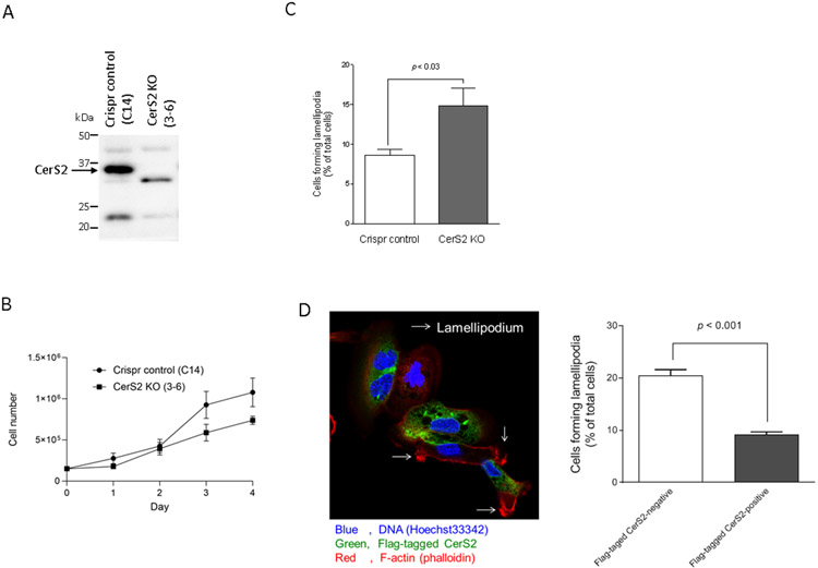
Figure 4. Effects of CerS2 knock-out and CerS2 knock-in on the formation of lamellipodia.
Cell lysates from CRISPR control (C14) and CerS KO (3-6) cells were examined by immunoblotting using antibodies specific to CerS2 (A). (B) CRISPR control (C14) and CerS KO (3-6) cells were plated on 6-well plates and incubated for up to 4 days. Cell numbers were determined by trypan blue staining (n = 6). (C) The formation of lamellipodia in CRISPR control (C14) and CerS KO (3-6) cells was assessed as described in “Materials and Methods” (n = 6). (D) CerS KO (3-6) cells were transfected with anti-Flag-tagged human CerS2 pCMVexSVneo vectors for 20 h. Cells were fixed followed by staining with Flag antibodies (Green), TRITC-conjugated phalloidin (red) and Hoechst 33342 (blue). Over 100 cells in each sample were assessed for negative- or positive-expression of Flag-tagged CerS2 and then the formation of lamellipodia was assessed as described in “Materials and Methods”. Data shown (mean ± standard error, n = 3) are the percentage of cells forming lamellipodia.

