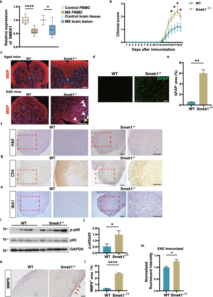
Fig. 1.
Suppressor of MEK1 (Smek1) reduction is related to multiple sclerosis (MS) and causes worsened experimental autoimmune encephalomyelitis (EAE) symptoms. a Box plot showing the transcriptional analysis of peripheral blood mononuclear cells from controls (n = 15), MS patients (n = 14), normal brain tissue (n = 3), and MS lesions (n = 9) from the Gene Expression Omnibus (GEO) dataset. PBMC, peripheral blood mononuclear cells. The box and whiskers are the median, minimum and maximum data for each group. Data are presented as the mean ± SEM and were analyzed by the two-sided unpaired t test; *P < 0.05; ****P < 0.0001. b Wild-type (n = 14) and Smek1-/+ (n = 17) mice were immunized with myelin oligodendrocyte glycoprotein 35-55 (MOG35-55) and CFA in 3 independent experiments. Daily clinical scores are shown as the mean ± SEM and were analyzed by the Mann-Whitney test, *P < 0.05. c Myelin basic protein (MBP) immunofluorescent staining of spinal cords from aged (10-month-old) mice and EAE mice. The white arrowheads indicate ruptured myelin; the yellow arrowheads indicate infiltrated cells (scale bar, 100 μm). d Immunofluorescent staining of spinal cords showing activated astrocytes (scale bar, 50 μm). e Quantification of GFAP staining in EAE spinal cords (n = 5 in each group). Data are presented as the mean ± SEM and were analyzed by the two-sided unpaired t test; **P < 0.01. f–h Hematoxylin-eosin (H&E) staining (e) and CD4 (f) and IBA1 (g) staining of spinal cord serial sections dissected from EAE spinal cords (scale bar, 100 μm). i Activation of the nuclear factor kappa B (NF-κB) signal detected by immunoblotting EAE brain tissue. j Western blot analysis was performed to evaluate the level of p-p65 (n = 8 in each group). Data are presented as the mean ± SEM and were analyzed by the two-sided unpaired t test; *P < 0.05. k Immunohistochemical staining of spinal cords for matrix metalloproteinase 9 (MMP9) levels. The red arrows point to dark stained tissue (scale bar, 50 μm). l Quantification of MMP9 deposition area in WT and Smek1-/+ EAE spinal cords (n = 5 in each group). Data are presented as the mean ± SEM and were analyzed by the two-sided unpaired t test; ****P < 0.0001. m Sodium fluorescein assay quantifying blood-brain barrier permeability in EAE mice (right) (n = 3 in each group) Data are presented as the mean ± SEM and were analyzed by the two-sided unpaired t test; *P < 0.05

