Fig. 4.
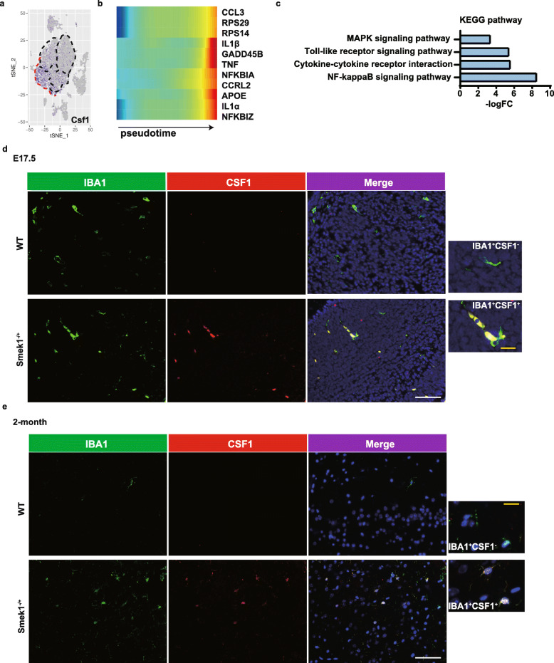
Proinflammatory colony stimulating factor 1 (Csf1)-positive microglia in the Smek1-deficient central nervous system (CNS). a tSNE plots of Csf1 expression in total microglia. b Heatmap of pseudotime gene expression in Smek1-/- microglia. c Kyoto Encyclopedia of Genes and Genomes (KEGG) analysis of marker genes in cluster 3. d, e Colony stimulating factor 1 (Csf1) staining in wild-type and Smek1-/+ mice at E17.5 (d) and 2 months old (e) (white scale bar, 25 μm). IBA1+Csf1- microglia in wild-type (upper) and IBA1+Csf1+ microglia in the Smek1-/+ cortex (lower) are magnified (yellow scale bar, 5 μm)

