
Figure 3. Effect of tetanus toxin microinjection on cortical granule exocytosis.
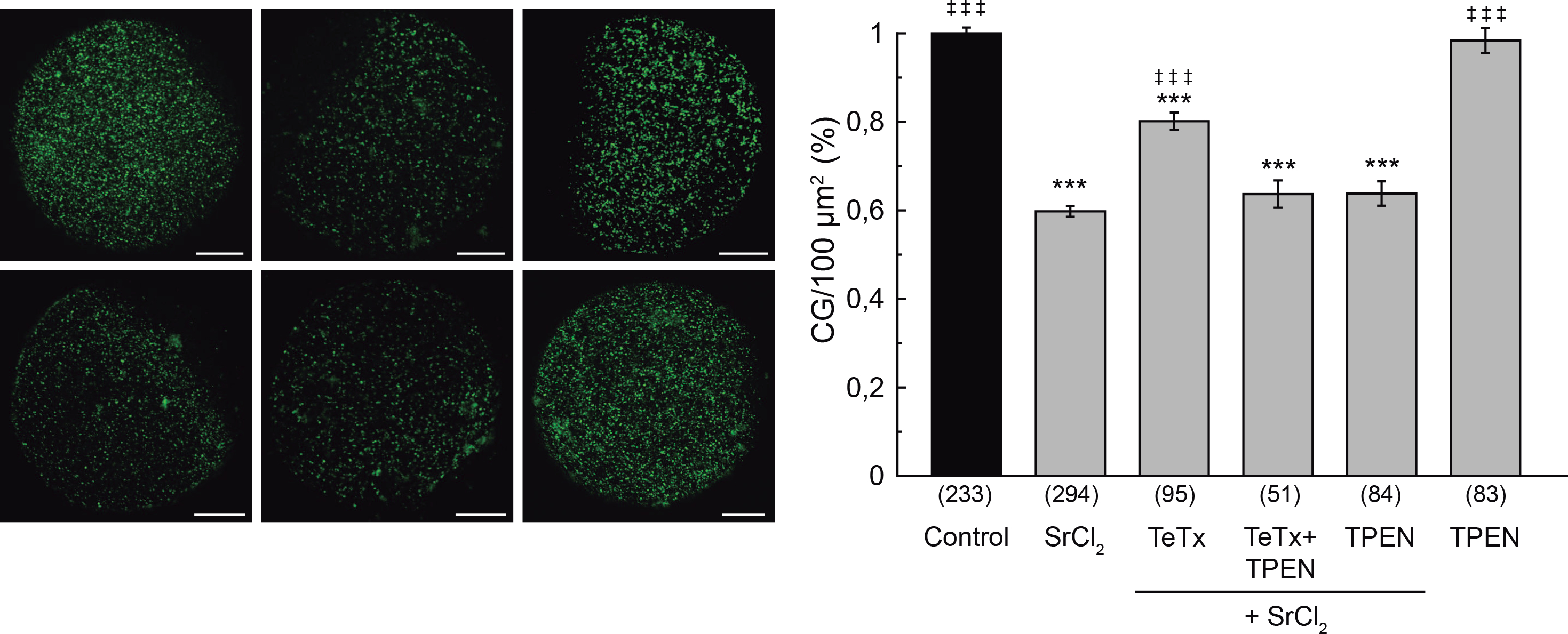
Oocytes were microinjected with 10 μM tetanus toxin (TeTx), and when indicated, incubated with TPEN 10 μM. Cortical granule exocytosis was triggered with 30 mM SrCl2. Images were taken as described in M&M. Left, representative confocal images of oocytes stained with FITC-LCA for each experimental condition. Scale bar: 20 μm. Right, histogram showing CG density/100 μm2 for different treatments, relative to untreated group (Control) set as 100%. Data are shown as mean ± SEM from 3 independent experiments. Numbers in parentheses below bars represent total number of oocytes. ***, values compared to control without stimulus, p ≤ 0,001; ‡ ‡ ‡, values compared to SrCl2, p ≤ 0,001. Statistical tests: One way ANOVA and Tukey’s test.