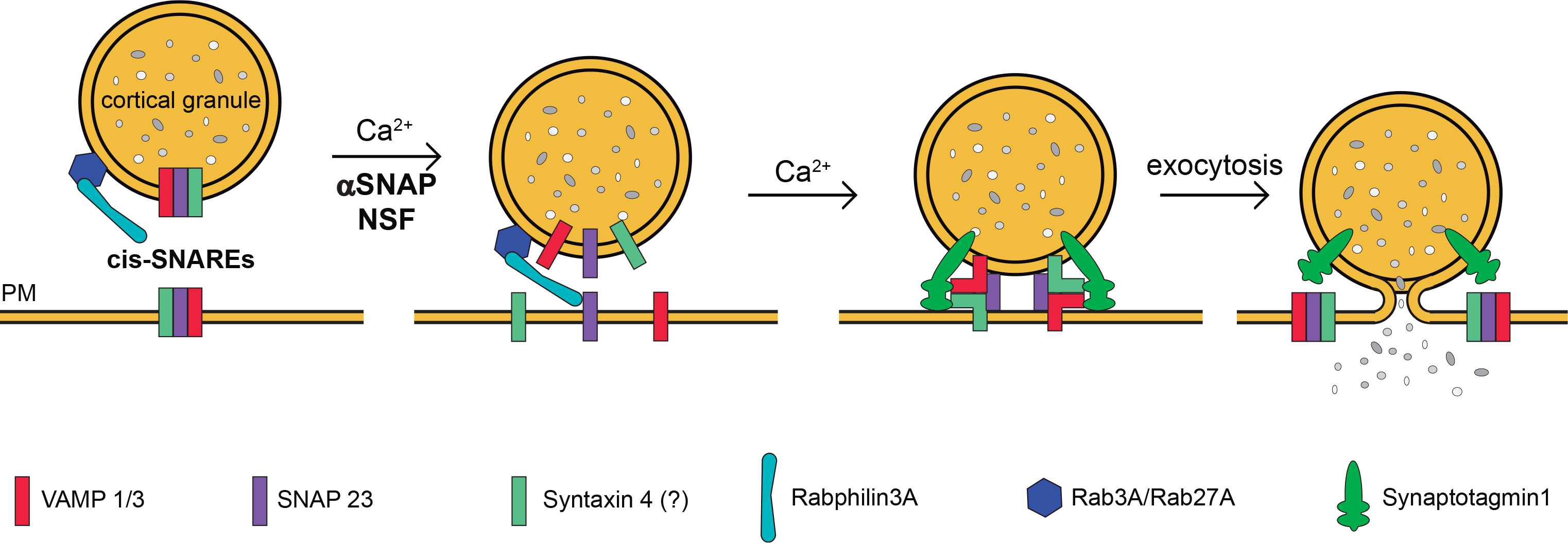
Figure 5. Working model for membrane fusion during cortical granule exocytosis in mouse oocytes.

Based on our previous and recent results and those published by other groups, we present this scheme to summarize the proteins that are involved in cortical granules exocytosis in mouse oocytes and their probable functions. We proposed that ternary cis-SNAREs complexes –formed by SNAP23 [34], Syntaxin4 [28] and VAMP1/3 (this work) are preformed on cortical granules (and probably on plasma membrane remnant from constitutive exocytosis). Rab3A-GTP [4], Rabphilin3A [32], and Rab27-GTP [54] are already recruited on cortical granules. During mouse oocyte activation, a sudden increase in the amount of calcium ions (Ca2+) would activate alpha-SNAP/NSF complex and Synaptotagmin1. Alpha-SNAP/NSF complex would disassemble preformed SNARE complexes [13] and would release their components to allow the fusion of vesicle granules and plasma membrane (PM). Meanwhile, Rabs/rabphilin complex would facilitate the tethering membranes. Finally, Synaptotagmin1[57] would detect calcium ions and would bind to the cell membrane and the new trans-SNAREs complexes allowing the secretion of cortical granule content.
