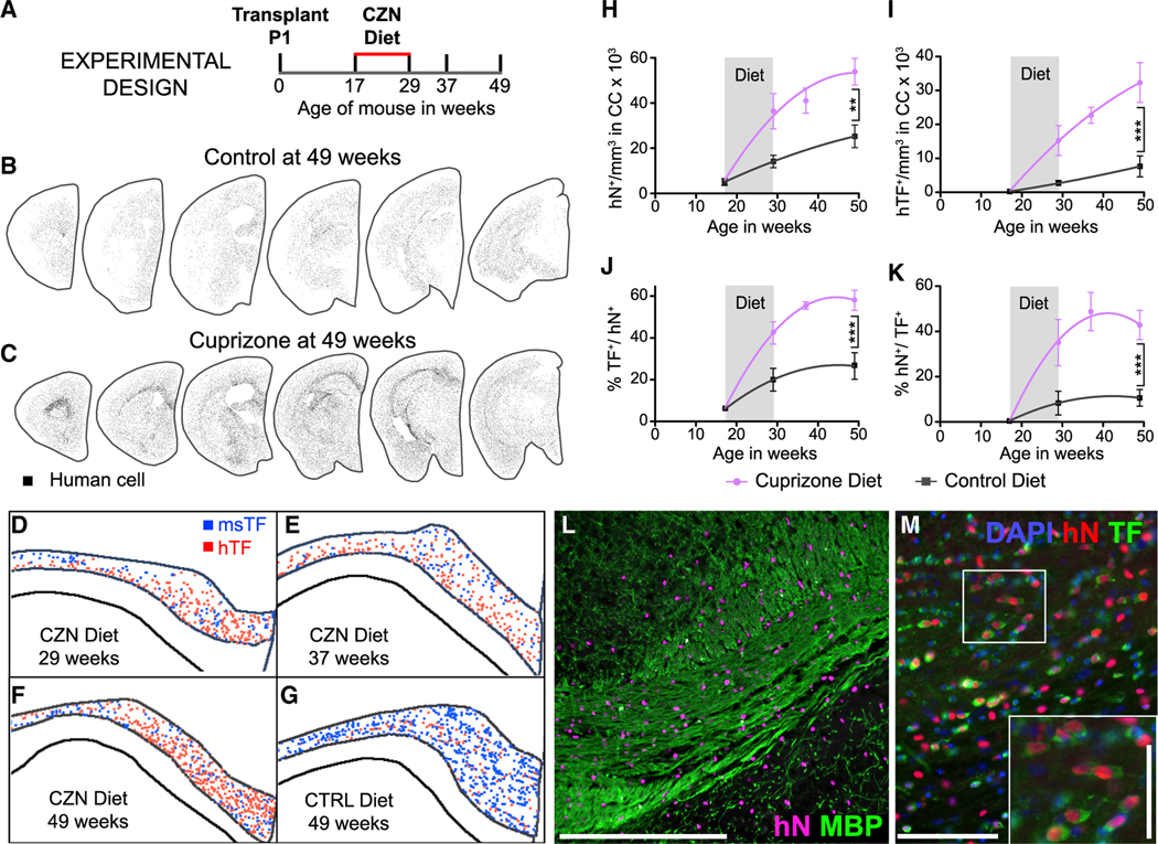
Figure 2. hGPCs Differentiate as Myelinogenic Oligodendroglia in Response to Cuprizone Demyelination.
(A) The schematic outlines the experimental design for neonatal engraftment followed by adult demyelination. Mice were transplanted with 2 × 10⁵ hGPCs perinatally, maintained on a control diet through 17 weeks of age, then placed on either a cuprizone-supplemented or normal diet for 12 weeks, then either sacrificed or returned to standard diet, and killed at later time points.
(B and C) Serial coronal sections comparing dot-mapped distributions of human (hNA) cells in control (B) and cuprizone-fed (C) mice at 49 weeks of age, following 20 weeks of recovery on the control diet.
(D–G) Relative positions and abundance of human (red dots) and mouse (blue) TF-defined oligodendrocytes, mapped in 20-mm coronal sections of corpus callosa of mice engrafted with hGPCs neonatally, demyelinated as adults from 17–29 weeks of age, and then assessed either at the end of the cuprizone diet (D), 8 weeks after return to control diet (E), or 20 weeks after cuprizone cessation (F). (G) Shows an untreated control, age-matched to (F).
(H) The density of human cells in the corpus callosum increases to a greater degree and more rapidly in cuprizone-demyelinated brains than in untreated controls, including during the 12-week period of cuprizone treatment (indicated in gray). **p < 0.001.
(I) By 8 weeks after the termination of cuprizone exposure, the density of human oligodendroglia was >5-fold greater in cuprizone-demyelinated than in untreated control brains. ***p = 0.0006.
(J and K) By that 80-week recovery point, over half of all hGPCs engrafted in the corpus callosa of cuprizone-treated mice had differentiated as oligodendrocytes (J), and accordingly, over half of all TF-defined callosal oligodendrocytes were human (K); in contrast, relatively few human oligodendrocytes were noted in untreated chimeric brains. ***p < 0.0001.
(L) Substantial colonization by human glia evident in this remyelinated corpus callosum, after a 20-week recovery period (human nuclear antigen, magenta; myelin basic protein, green).
(M) Chimeric white matter populated, after cuprizone demyelination, by hGPC-derived oligodendroglia. hNA, red; TF, green; inset highlights relative abundance ofhNA+/TF+ human oligodendroglia. Scale bars: 100 μm (L) and 50 μm (M); inset scale bar: 25 μm.

