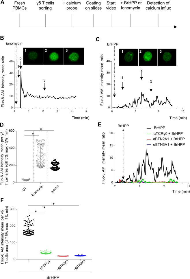
Fig. 2.
Self-activation of resting purified Vγ9Vδ2 T cells by exogenous BrHPP is dependent on TCR, BTN3A1, and BTN2A1. A Sequence of actions for calcium flux detection by video in an individual Vγ9Vδ2 T cell. B–E Time lapse of the Fluo-8 AM intensity representing the calcium flux in one Vγ9Vδ2 T-cell stimulated by ionomycin (B) or exogenous BrHPP (C). Three images were extracted from the time lapses at three different time points of the stimulation. D Mean Fluo-8 AM intensity per γδ T cell area (difference 5% max–5% min). F Time lapse of the Fluo-8 AM intensity representing the calcium flux in one Vγ9Vδ2 T-cell stimulated by BrHPP in the presence or absence of blocking antibodies against γ9TCR, BTN3A1. or BTN2A1. Asterisk (*) indicates p < 0.05, Student’s paired t test; ns not significant

