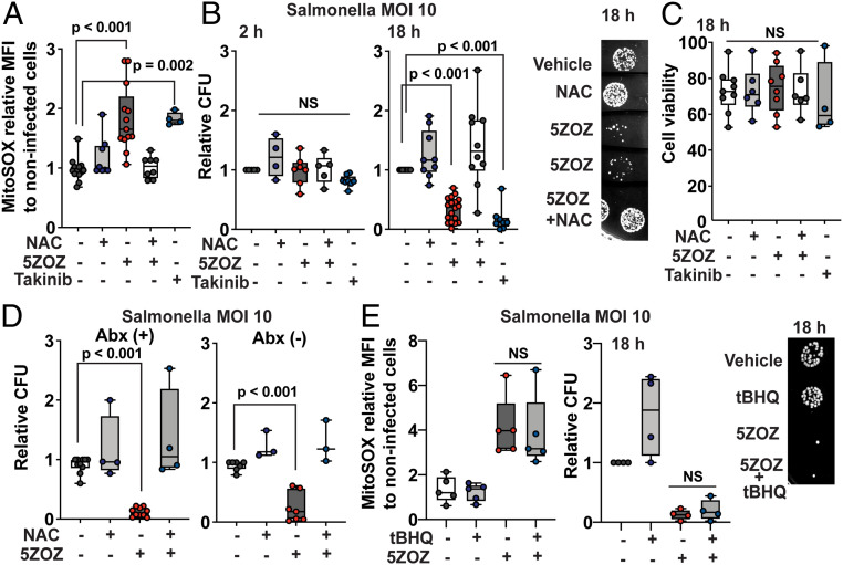
Fig. 3.
TAK1 inhibition blocks intracellular Salmonella growth through mitochondrial ROS. (A and B) BMDMs were incubated with Salmonella, MOI of 10, for 30 min, and extracellular Salmonella was killed by gentamicin treatment. TAK1 inhibitors, 300 nM 5ZOZ and 10 µM Takinib, and 3 mM NAC were added to the medium when starting gentamicin treatment. Cells were analyzed at 18 h post infection for ROS (A). The median intensity of live cells relative to that in noninfected cells with vehicle treatment is shown. Intracellular bacteria numbers were determined at 2 and 18 h post infection (B). Representative Salmonella colonies from cell lysate at 18 h are shown (B, Right). (C) Cell viability was assessed by SytoxGreen staining at 18 h post infection. (D) BMDMs were infected with Salmonella using the same procedure of A and B. At 2 h of gentamicin treatment, culture medium was changed to the same medium with gentamicin (left graph) or to a medium without antibiotics (right graph). (E) BMDMs were infected with Salmonella using the same procedure of A and B. A general ROS scavenger, 20 µM tBHQ, was cotreated with 300 nM 5ZOZ. Representative Salmonella colonies from cell lysate at 18 h post infection are shown (Right). One-way ANOVA, multiple comparisons, and Tukey test; N.S., not significant.

