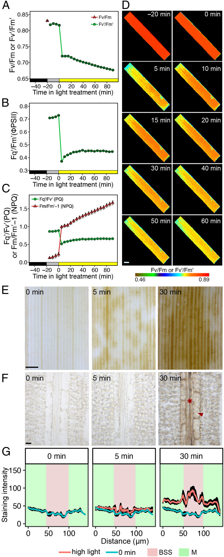
Fig. 1.
Rice BSS preferentially accumulate the DAB polymerization product in response to high light. (A–C) Chlorophyll fluorescence parameters associated with dark-adapted leaves being moved into the light intensity of growth for 20 min and then moved to a 10-fold higher intensity of light. (A) Dark-adapted Fv/Fm and Fv’/Fm’. (B) Quantum efficiency of Photosystem II (Fq’/Fm’ or ΦPSII) and (C) PQ and NPQ. Data shown represent mean and SE from 16 leaves. (D) Representative images from the chlorophyll fluorescence imager showing responses were reasonably homogenous across the leaf. (Scale bar: 2 mm.) (E) High-light stress led to strong staining from the DAB polymerization product in BSS arranged along the proximal to distal axis of the leaf blade. After 5 min of high light, staining is evident, but at 30 min, it is stronger and more homogenous in these BSS. (Scale bar: 1 mm.) (F) Representative image from paradermal sections show that cells accumulating DAB stain are veins (asterisk) and bundle-sheath cells (arrowhead). (Scale bar: 10 μm.) (G) Semiquantitation of DAB stain in M cells and BSS. Data are presented as mean (red or blue line) and one SE from the mean, n = 4).

