Fig. 6.
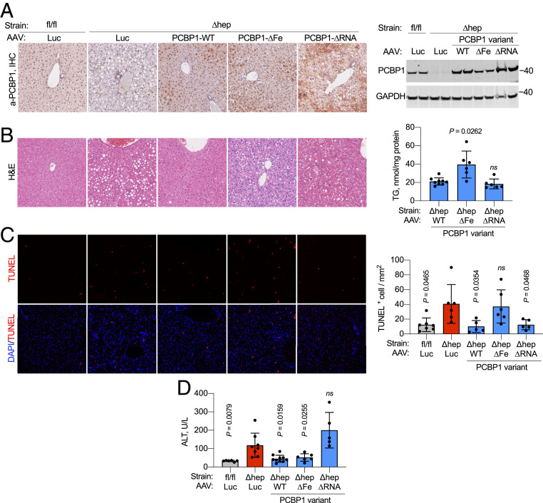
RNA- and iron-binding activities of PCBP1 are associated with different phenotypes in PCBP1-depleted hepatocytes. (A) Transduction of AAV constructs with PCBP1 WT, RNA-binding mutant (∆RNA), or iron-binding mutant (∆Fe) in PCBP1 WT (fl/fl) and deleted (∆hep) livers. (Left) Representative immunohistochemistry showing the distribution of PCBP1 in AAV-transduced livers. AAV-luciferase (Luc) was used as negative control. Shown is a representative immunoblot of PCBP1 in liver lysates from PCBP1fl/fl and PCBP1 ∆hep mice injected with the indicated AAV constructs. (Right) GAPDH used as a loading control. (B) Persistence of steatosis in livers transduced with PCBP1 ∆Fe. (Left) Hematoxylin and eosin (H&E) staining of PCBP1fl/fl and PCBP1 ∆hep mice injected with AAVs. (Right) Total acylglycerols (TG) in livers of PCBP1∆hep mice injected with the PCBP1 variant AAVs. (C) Increased TUNEL+ cells in liver from PCBP1 ∆hep mice and PCBP1 ∆hep mice expressing ∆Fe mutant. Representative images of liver sections stained with TUNEL and DAPI (Left) with quantitation (Right). (D) RNA-binding activity of PCBP1 is required to suppress hepatocyte cell death. Plasma ALT levels as units per liter (U/L) in PCBP1fl/fl and PCBP1 ∆hep mice transduced with AAVs. Data represent means ± SD; P values calculated using one-way ANOVA and unpaired t test with Welch’s correction. ns, not significant. See also SI Appendix, Fig. S6.

