Fig. 1.
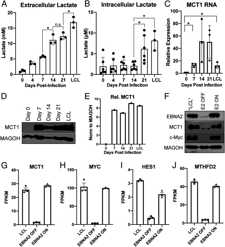
EBV infection of human B cells leads to an increase in lactate transport through MCT1. (A) Extracellular (n = 3) and (B) Intracellular (n = 6) lactate concentration during EBV-infected B cell immortalization. Extracellular concentration was obtained from the supernatant of CD19+-isolated B cells at a concentration of 1 × 10⁶/mL and corrected to cell-free growth medium lactate concentrations. Intracellular lactate concentration is per 12,500 cells. (C) MCT1 RNA (n = 3) and (D) protein levels during EBV-infected B cell immortalization. MAGOH is a loading control used because its expression does not change during B cell outgrowth. (E) Quantitation of MCT1 protein levels from D, normalized to MAGOH. (F) Immunoblot of EBNA2, MCT1, and c-Myc in P493-6 cells at the “LCL” state with stable EBNA2 expression, EBNA2 OFF in the absence of β-estradiol, and EBNA2 ON with EBNA2 expression regained. (G–J) RNA-seq data showing MCT1, c-Myc, HES1, and MTHFD2 expression in P493-6 B cells (n = 3). FPKM = fragments per kilobase of transcript per million mapped reads. All statistical significance was determined by a paired Student’s t test, in which *P < 0.05 and n.s. (not significant) is P ≥ 0.05.

