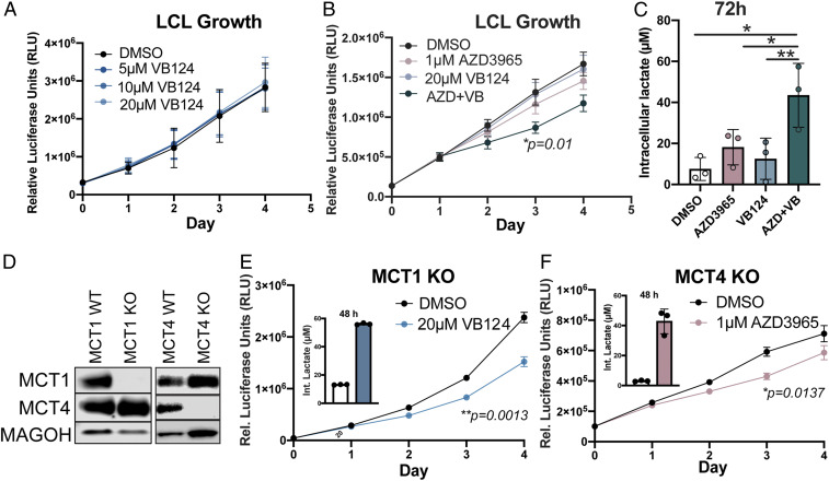
Fig. 4.
Dual MCT1/4 inhibition reduces LCL growth and increases intracellular lactate. (A) Growth curve of LCLs (n = 3) treated with varying concentrations of VB124. Growth was determined by CellTiter Glo. (B) Growth curve of LCLs (n = 3) treated with either DMSO, 1 µM AZD3965, 20 µM VB124, or 1 µM AZD3965 + 20 µM VB124. Growth was assessed as in A. Statistical significance was determined for day 3 of treatment. (C) Intracellular lactate concentration (per 12,500 cells) at 72 h of LCLs (n = 3) treated with either DMSO, 1 µM AZD3965, 20 µM VB124, or 1 µM AZD3965 + 20 µM VB124. (D) Western blot representation of CRISPR-Cas9–mediated MCT1- and MCT4-knockout LCLs. MAGOH = loading control. (E) MCT1-knockout LCL growth (n = 3) over time and intracellular lactate concentration at 48 h following treatment with 20 µM VB124. Statistical significance was calculated at day 3. (F) MCT4-knockout LCL growth (n = 3) over time and intracellular lactate concentration at 48 h following treatment with 1 µM AZD3965. Statistical significance was calculated at day 3. Statistical significance was determined by paired Student’s t test in which *P < 0.05 and **P < 0.01.

