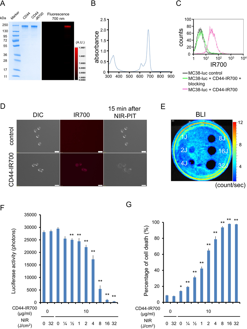
Figure 1. Confirmation of CD44 expression as a target for NIR-PIT in MC38-luc cells and evaluation of in vitro NIR-PIT.
(A) Validation of CD44-IR700 by SDS-PAGE (left: Colloidal Blue staining; right: IR700 fluorescence). Diluted anti-CD44 was used as a control. (B) Absorbance curve of CD44-IR700. (C) Expression of cell surface CD44 in MC38-luc cells was examined with flow cytometry. CD44-blocking antibody was added to some wells to validate specific staining. Representative histograms shown. (D) Differential interference contrast (DIC) and fluorescence microscopy images of MC38-luc cells. Change in MC38-luc cellular architecture following 15 minutes of NIR light exposure shown. Scale bars = 20 μm. (E) Bioluminescence imaging (BLI) demonstrating luciferase activity in MC38-luc cells following NIR-light. (F) Quantification of MC38-luc luciferase activity after labelling with CD44-IR700 and treatment with NIR-light (n = 5, **p < 0.01 vs. untreated control, Student’s t test). (G) Membrane permeability of MC38-luc cells, as measured by propidium iodide (PI) staining, after labeling with CD44-IR700 and treatment with NIR-light (n = 5, *p < 0.05 vs. untreated control, **p < 0.01 vs. untreated control, Student’s t test). Each value represents mean ± standard error of the mean (SEM) of five independent experiments.

