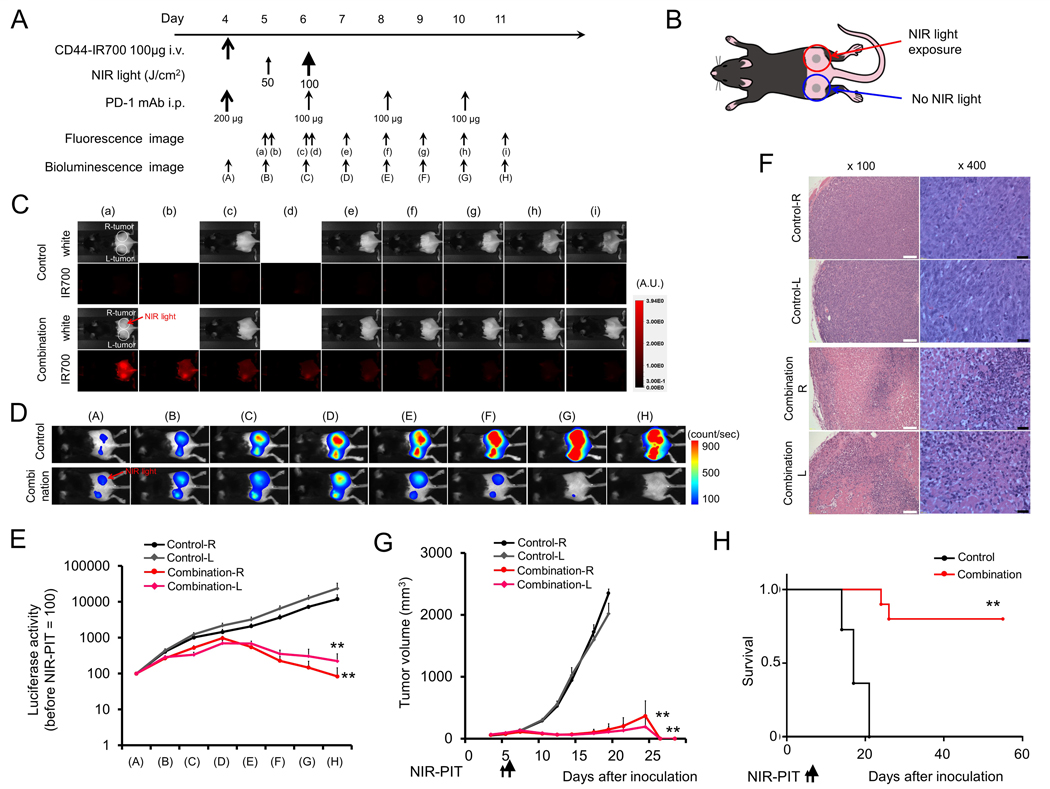
Figure 4. In vivo effect of NIR-PIT and PD-1 mAb in mice bearing bilateral MC38-luc tumors.
(A) NIR-PIT regimen. Bioluminescence and fluorescence images were obtained at each time point as indicated. (B) Light exposure. NIR light was administered to the right-side tumor only in mice bearing bilateral lower flank tumors. The untreated left-side tumor was shielded from NIR light. (C) In vivo IR700 fluorescence real-time imaging of tumor-bearing mice in response to NIR-PIT to the right-side tumor only. (D) In vivo BLI of tumor bearing mice in response to combination NIR-PIT and PD-1 mAb. (E) Quantification of luciferase activity from each tumor in controls and mice treated with combination NIR-PIT and PD-1 mAb (n = 10, **p < 0.01, Tukey’s test with ANOVA). (F) Resected tumors (day 10) were stained with H&E and assessed for necrosis and leukocyte infiltration. White scale bars = 100 μm. Black scale bars = 20 μm. (G) Growth curves of right- and left-side tumors from controls and mice treated with combination NIR-PIT and PD-1 mAb. (H) Kaplan-Meier survival analysis from controls and mice treated with combination NIR-PIT and PD-1 mAb (n = 10, **p < 0.01, Tukey’s test with ANOVA for growth curves; **p < 0.01, Log-rank test for survival).

