
Figure 1.
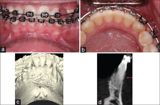
Preoperative frontal (a) and lingual (b) views. Note presence of c d slight marginal erythematous tissues localized to the mandibular arch. Note the thin biotype with scalloped gingival margins and a limited width of keratinized tissue that ranges from 2 to 3 mm from #23 to 26. Preoperative cone-beam computerized tomography scan showing lingual view of the mandibular incisors (c), and a thin mandibular labial bone on the transaxial section for tooth #22 (d)