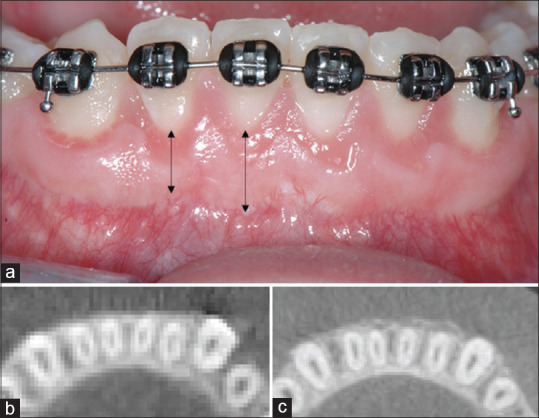
Figure 5.

(a) Frontal view of the buccal aspect of the mandibular anterior teeth 2.1-year postoperatively to periodontally accelerated osteogenic orthodontics. Note the presence of stable band of keratinized tissue that ranged from 4 to 6 mm. (b) Preoperative and (c) 2.1 years' postperiodontally accelerated osteogenic orthodontics cone beam computerized tomography axial section. Note presence buccal bone increased on the mandibular anterior teeth (b), where prior to periodontally accelerated osteogenic orthodontics was absent (c)
